Airway morphological parameter quantitative acquisition method and device and airway stent design method
An acquisition method and morphological technology, which is applied in the field of respiratory morphological parameter measurement, can solve the problems of inaccurate and irregular quantitative measurement of bronchial tree morphological parameters, and achieve the effect of reducing complications and accurate quantitative parameters
- Summary
- Abstract
- Description
- Claims
- Application Information
AI Technical Summary
Benefits of technology
Problems solved by technology
Method used
Image


Examples
Embodiment 1
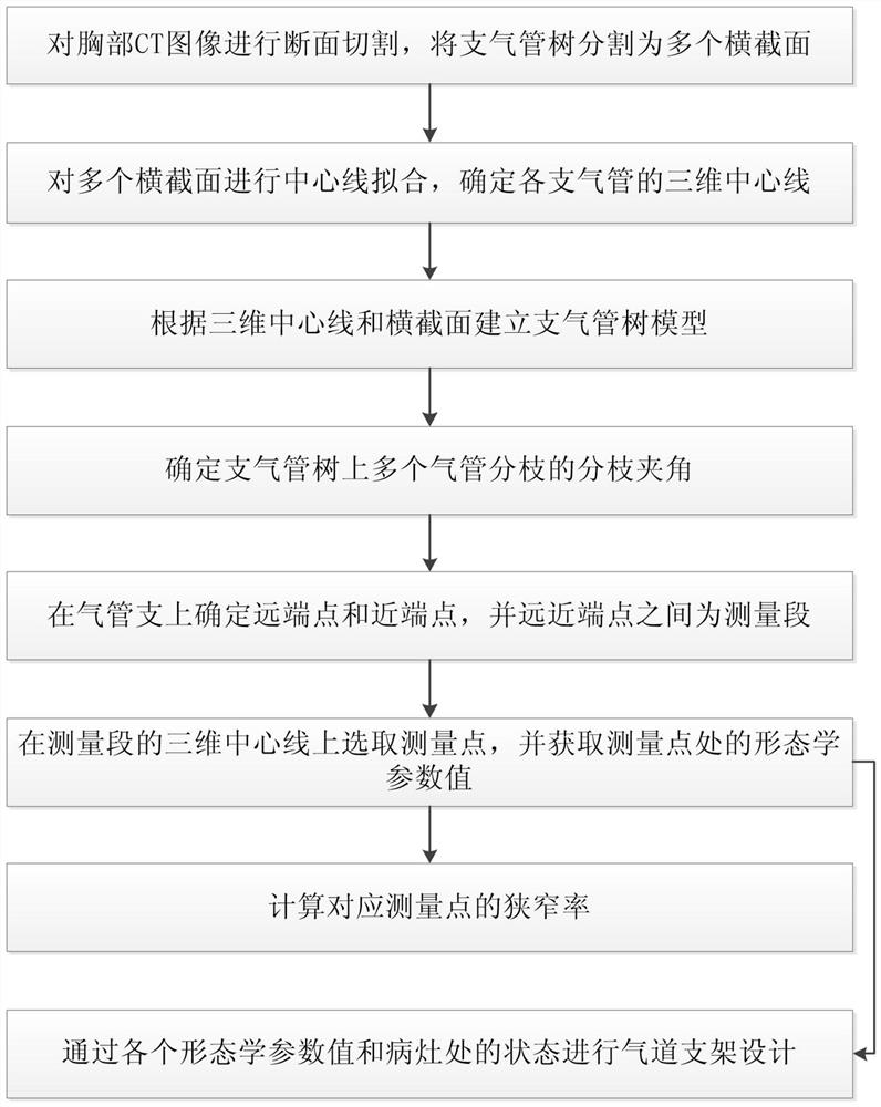
[0071] In view of the above defects, this embodiment proposes a method for quantitatively acquiring airway morphological parameters, including the following steps:
[0072] The first step is to section the chest CT image, and divide the bronchial tree into multiple cross-sections; in the section section of the chest CT image, the segmentation method adopts sub-pixel segmentation accuracy, which can ensure the accuracy of tracheal segmentation.
[0073] The second step is to fit the centerlines of multiple cross-sections to determine the three-dimensional centerlines of each bronchi, that is, to first determine the center points of each section, and connect the center points in turn to form the center line of the bronchial tree.
[0074] The third step is to establish a bronchial tree model according to the three-dimensional centerline and cross-section, and build a model of the bronchial tree in the modeling software according to the structure of the cross-section and the path of
Embodiment 2
[0111] Based on a quantitative acquisition method of airway morphological parameters in Example 1, after measuring various quantitative parameters such as the inner diameter of the bronchial tree, the area of the lumen, the length of the lesion, and the included angle of the branches, an auxiliary method is carried out according to the above data. For the design of the airway stent, the present embodiment provides an auxiliary airway design method based on the quantitative parameters of the airway, comprising the following steps:
[0112] Determine the lumen diameter, lumen area, lesion length, lumen volume, and branch angle corresponding to the lesion site by the method described in the article;
[0113] The inner diameter of the airway stent corresponds to the inner diameter of the lumen, the area of the stent corresponds to the area of the lumen, the length of the stent corresponds to the length of the lesion, the volume of the stent corresponds to the volume of the lumen
Embodiment 3
[0119] This embodiment provides a device for quantitatively acquiring airway morphological parameters in Embodiment 1, including a cutting module, a fitting module, a modeling module, a measurement module, a research area selection module, a first calculation module and Second computing module.
[0120] The cutting module is used for cross-sectional cutting of chest CT images, and the bronchial tree is divided into multiple cross-sections;
[0121] The fitting module is used for performing centerline fitting on multiple cross-sections to determine the three-dimensional centerline of each bronchi;
[0122] a measurement module for determining branch angles of multiple tracheal branches on the bronchial tree;
[0123] The research area selection module is used to determine the distal point and the proximal point on the bronchial tree, and select the bronchi between the distal point and the proximal point as the measurement segment;
[0124] The first calculation module is used to
PUM
 Abstract
Description
Claims
Application Information
 - R&D Engineer
- R&D Manager
- IP Professional
- Industry Leading Data Capabilities
- Powerful AI technology
- Patent DNA Extraction
Browse by: Latest US Patents, China's latest patents, Technical Efficacy Thesaurus, Application Domain, Technology Topic.
© 2024 PatSnap. All rights reserved.Legal|Privacy policy|Modern Slavery Act Transparency Statement|Sitemap