Miniature Glaucoma Shunt
- Summary
- Abstract
- Description
- Claims
- Application Information
AI Technical Summary
Benefits of technology
Problems solved by technology
Method used
Image

Examples
Example
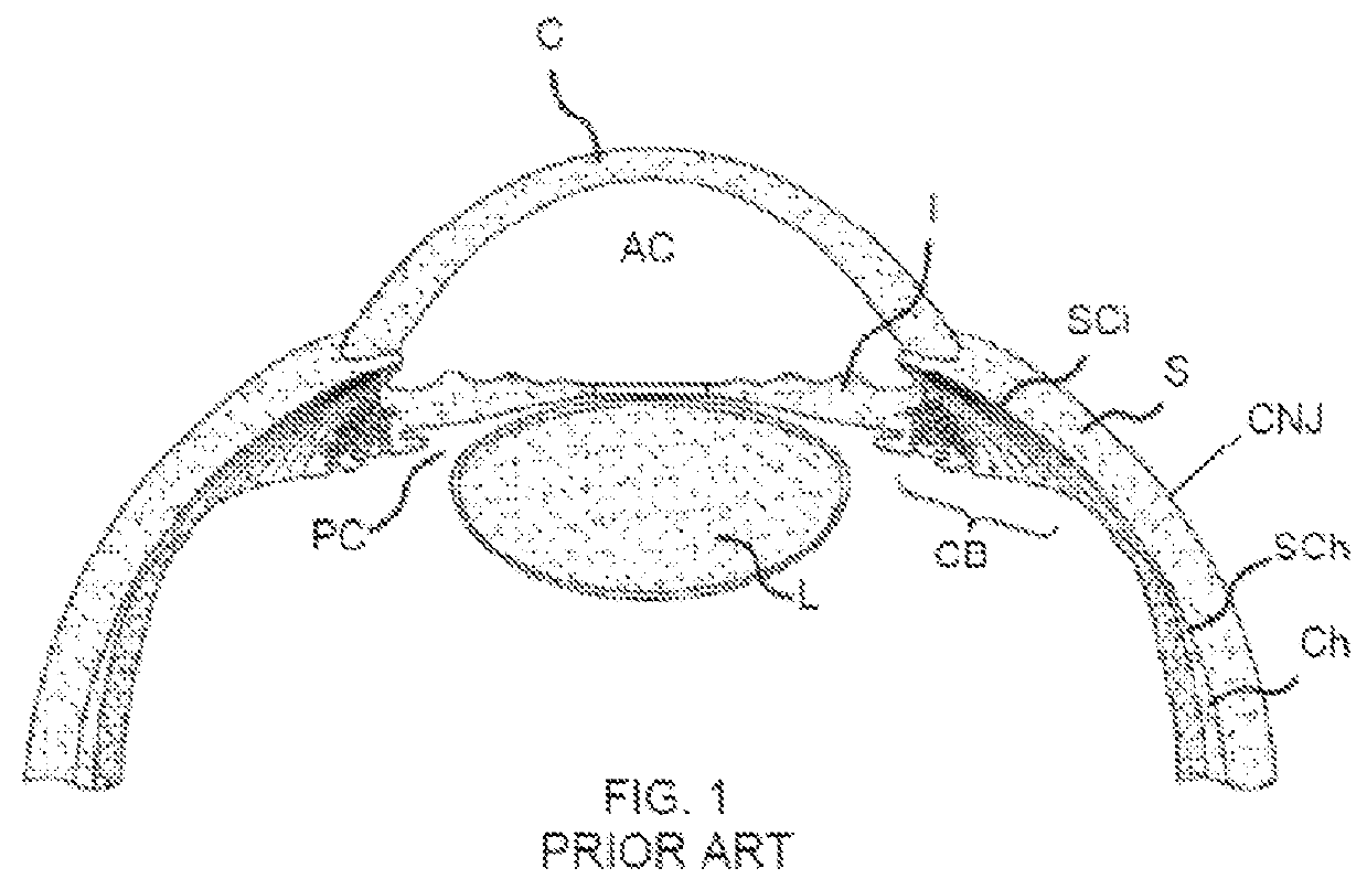
[0018]Reference is made to FIG. 1, which illustrates the anatomy of the human eye, and is presented here for better understanding of the implantation of the glaucoma shunt of the present invention.
[0019]The eye is covered on the outside by the sclera S and the cornea C. The conjunctiva CNJ lines the inside of the eyelids and covers the sclera S. The lens L is located near the front of the eye. The lens L provides adjustment of focus and is suspended within a capsular bag from the ciliary body CB, which contains the muscles that change the focal length of the lens. A volume in front of the lens L is divided into two by the iris I, which controls the aperture of the lens and the amount of light striking the retina. The pupil is a hole in the center of the iris I through which light passes. The volume between the iris I and the lens L is the posterior chamber PC. The volume between the iris I and the cornea C is the anterior chamber AC. Both chambers are filled with aqueous humor, a clear
PUM
 Abstract
Description
Claims
Application Information
 - R&D Engineer
- R&D Manager
- IP Professional
- Industry Leading Data Capabilities
- Powerful AI technology
- Patent DNA Extraction
Browse by: Latest US Patents, China's latest patents, Technical Efficacy Thesaurus, Application Domain, Technology Topic.
© 2024 PatSnap. All rights reserved.Legal|Privacy policy|Modern Slavery Act Transparency Statement|Sitemap