Auxiliary examination device for otolaryngology department

An otolaryngology and auxiliary device technology, applied in otoscopes, rhinoscopes, laryngoscopes, etc., can solve problems such as difficult inspections, inspection and diagnosis errors, etc., achieve high inspection efficiency, good accuracy, and improve inspection comfort Effect

Inactive Publication Date: 2019-04-19
佳木斯大学附属第二医院
View PDF5 Cites 1 Cited by
  • Summary
  • Abstract
  • Description
  • Claims
  • Application Information

AI Technical Summary

Benefits of technology

This new technology allows medical professionals inspect ears or noses more easily without having them bump into each other during examinations. It achieves these benefits through a device that expands outwardly on one side when used along certain directions. Additionally, there's another feature where the camera itself has a special lens system designed to capture images better than traditional cameras. These improvements make imagery taken from any view inside the body easier even after multiple shots have been done. Overall, they improve image quality and reduce stress caused due to prolonged exposures compared to previous methods.

Problems solved by technology

Technological Problem: Current methods used by physicians during otoacupuncture are time-consuming and require multiple steps involving various instruments such as xenon lamps that expose patients’ ears through direct light exposures. Additionally, there has been no effective way to inspect deeper areas within these holes without making them invisible due to their proximity to sensitive parts like eyes.

Method used


Image

  • Auxiliary examination device for otolaryngology department
  • Auxiliary examination device for otolaryngology department
  • Auxiliary examination device for otolaryngology department

Examples


Embodiment Construction

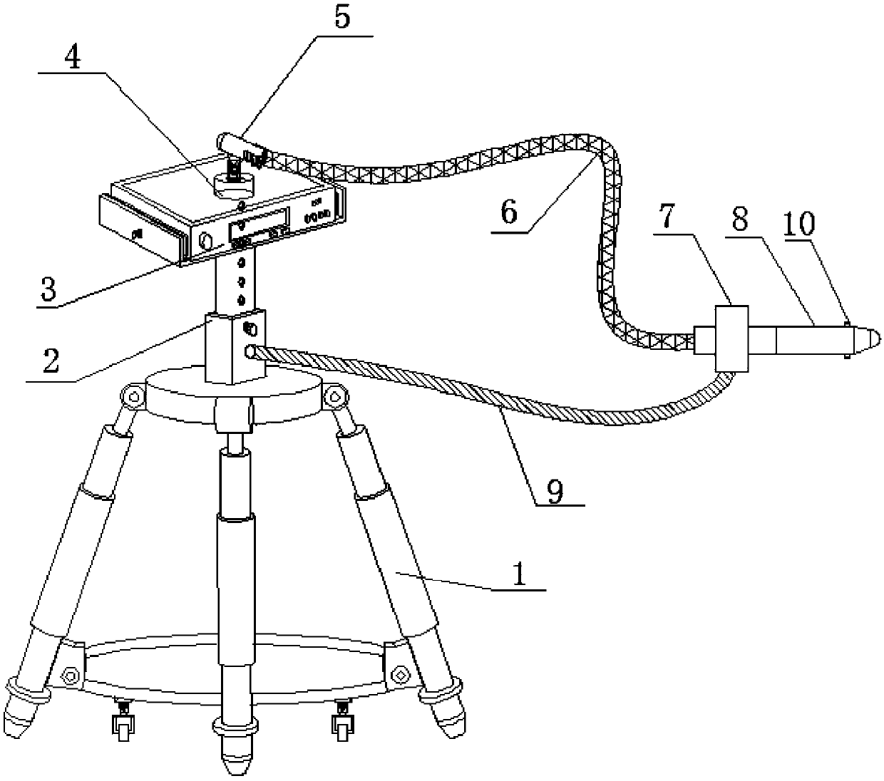
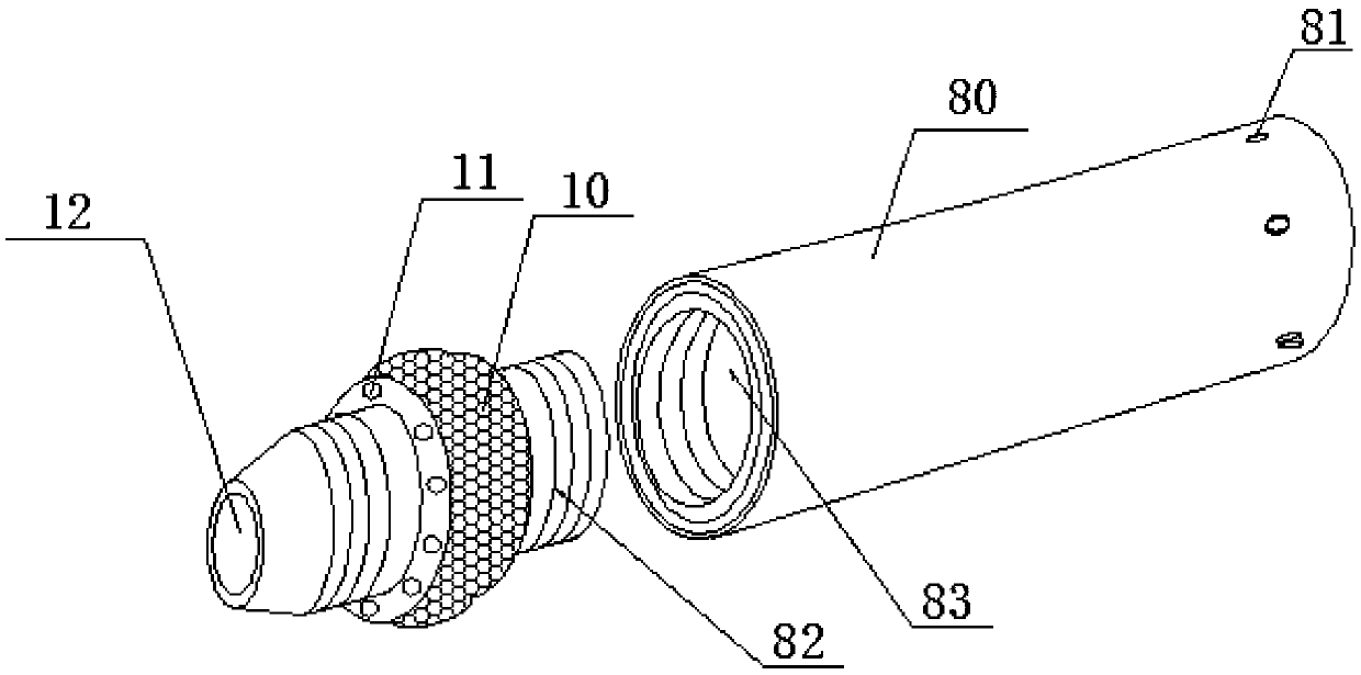
[0026] see Figure 1~5 , in the embodiment of the present invention, the auxiliary device for ENT examination includes an installation chassis 1, a telescopic support member 2, a support frame, a clamping fixing assembly 7 and an expansion angle adjustment mechanism 10, wherein the installation chassis 1 The top adopts a telescopic support member 2 to adjust the height of the supporting installation and is provided with the support frame, the inspection instrument 3 is fixedly installed on the support frame, and the inspection instrument 3 is connected with an inspection head that can be inserted into the ear or inside the nose through the control transmission line 6 8; characterized in that,

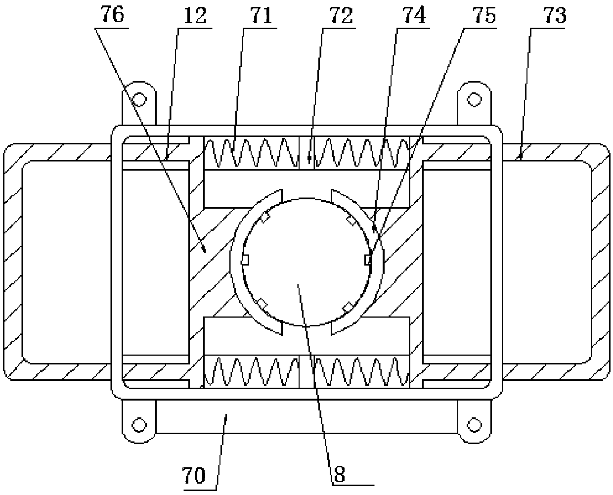
[0027] The inspection head 8 is detachably clamped on the clamping and fixing component 7, and the clamping and fixing component 7 is supported by an adjustment support 9, and the adjustment support 9 is designed to be flexible and deformable, so as to pass Its flexible deformation realiz


PUM


Abstract

The invention discloses an auxiliary examination device for the otolaryngology department. When the device is used for examination, the outer portion of the front end of an examination head is sleevedwith an expansion angle adjusting mechanism, and in this way, through the mode of expanding at all angles, the expansion angle adjusting mechanism can extrude the skin of the examination part at different angles to achieve video examination at all the angles; meanwhile, through the cooperation of the illumination of an illumination lamp and the design of a transparent structure, the internal conditions of the ears and nose can be clearly examined, the examination efficiency is high, the accuracy is good, and the comprehensive examination of various parts can be achieved. According to the auxiliary examination device, the examination head is supported by an adjusting support piece with a flexible deformable design, the angle of the examination head can be adjusted, positioning accuracy andreliability are ensured while the two hands of a doctor are freed, and meanwhile the examination comfort of an examined patient is improved.

Description


Claims


Application Information

Patent Timeline
Owner 佳木斯大学附属第二医院
Why Eureka
Try Eureka