Cerebrovascular image segmentation method based on statistical model and multi-scale filtering

An image segmentation and multi-scale technology, applied in image analysis, neural learning methods, image enhancement, etc., can solve the problem of poor segmentation effect of low-intensity small blood vessel images, and achieve the effect of improving segmentation accuracy

Pending Publication Date: 2022-03-01
BEIJING INSTITUTE OF TECHNOLOGYGY
View PDF0 Cites 2 Cited by
  • Summary
  • Abstract
  • Description
  • Claims
  • Application Information

AI Technical Summary

Benefits of technology

This patented describes two methods used during medical imaging: 1) by extracting specific features that help distinguish between different types or areas within an area being imaged (such as arteries), 2) by combining these extracted feature values into one more complex mathematical equation called the regression analysis algorithm, resulting in improved models over time. These techniques aim to enhance the quality of images obtained through optics technology while reducing errors caused by factors like shading or partial occlusion.

Problems solved by technology

This patented technical problem addressed in this patents relates to improving understanding how complex necrotic strokes (catheterization) affect the structure and behavior of nerves involved in the body during cardiovascuum events like heart attacks and strokes caused by blockages from arteries. Current techniques only involve analyzing entire regions through manual inspections, making it difficult to identify tiny blebs within those areas where they may develop complications related to reduced flow rates or increased risk of embolism.

Method used


Image

  • Cerebrovascular image segmentation method based on statistical model and multi-scale filtering
  • Cerebrovascular image segmentation method based on statistical model and multi-scale filtering
  • Cerebrovascular image segmentation method based on statistical model and multi-scale filtering

Examples


Embodiment 1

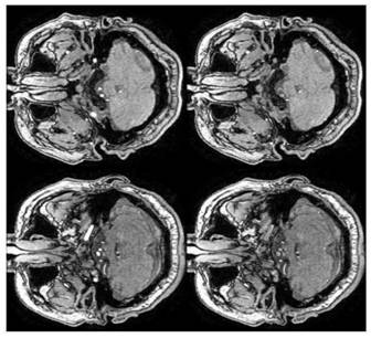
[0100] Cerebrovascular disease is one of the main diseases leading to human death, and its early prevention and treatment are extremely important. Non-invasive detection of cerebrovascular lesions by imaging technology is an effective method for diagnosing and monitoring cerebrovascular diseases, and it is also the most acceptable technology without side effects. This research provides technical support for the diagnosis and prevention of cerebrovascular diseases, and related technologies can be extended to other fields such as multimodal image registration and 3D visualization. Due to the large number of people who participate in physical examination and preventive health care every year in our country, the application of this research result can produce extensive social and economic benefits.

[0101] In this example, the Brave data set is used as our target object; Brave, Brain and VascularHealth in the Elderly, the purpose of this data set research is to use multimodal MRI to


PUM


Abstract

The invention provides a cerebrovascular image segmentation method based on a statistical model and multi-scale filtering, and belongs to the technical field of medical calculation and image processing. The segmentation method combines a statistical model and multi-scale filtering so as to combine a high-level model and a low-level model to extract a cerebrovascular image and segment the cerebrovascular image, and comprises the following steps: S1, preprocessing data to obtain a denoised brain region image; s2, fitting a Gaussian model of an image background and a blood vessel class in the S1, and building an FMM; s3, setting an FMM initial value, estimating parameters and calculating a low-layer energy function; s4, segmenting the image; and S5, screening the seed points in combination with ridge line properties, extracting a blood vessel center line in a 3D space, performing expansion along the far-end blood vessel center line, and supplementing the low-layer model. According to the method, the segmentation precision of the far-end blood vessel image is improved, and accurate blood vessel center line extraction can be achieved under a rough blood vessel image segmentation result.

Description


Claims


Application Information

Patent Timeline
Owner BEIJING INSTITUTE OF TECHNOLOGYGY
Why Eureka
Try Eureka