Ultrasonic diagnostic device and image information management device
An image information and diagnostic device technology, applied in the directions of sonic diagnosis, infrasound diagnosis, ultrasonic/sonic/infrasonic diagnosis, etc., can solve problems such as troublesome interpretation of doctors
- Summary
- Abstract
- Description
- Claims
- Application Information
AI Technical Summary
Problems solved by technology
Method used
Image

Examples
Embodiment 2
[0097] Embodiment 2 will describe a case where output information is generated only in specified ultrasonic images.
[0098] The output information generating unit 18 related to the second embodiment generates output information only in the ultrasonic image designated by the operator. For example, the operator designates whether to use the composite image for outputting output information when saving the composite image. Accordingly, the image combining unit 16 associates and stores the ultrasonic image together with the first image and a flag for output, for example, in the image memory 15 . Furthermore, when the output information generation unit 18 according to the second embodiment has accepted the output request of the output information of the ultrasonic examination of the subject P, among the plurality of ultrasonic images of the subject P, only the flags are associated with each other. Ultrasound image to generate the output image.
[0099] Second, use Figure 14 , the
Embodiment 3
[0104] In Example 3, for the processing using the output information, use Figure 15 Be explained. Figure 15 It is a figure for explaining the control part concerning Example 3.
[0105] The control unit 19 related to the third embodiment controls to display on the monitor 2 an ultrasonic image from which the specified image information is extracted when the operator who refers to the output information specifies the image information included in the output information. .
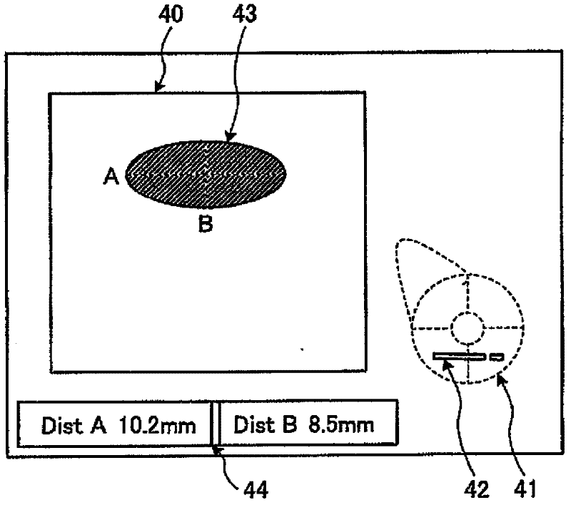
[0106] Below, use the Figure 8 The output information 57 described above describes the processing of the control unit 19 related to the third embodiment. For example, if Figure 15 As shown in the left figure of , when the interpreter designates the image 55 of the measurement target site schematically showing the measurement result "Φ(phi) 10.2×8.5" through the mouse of the input device 3, the control unit 19 The relative position information of 55 is read out from the image memory 15 as a composite im
PUM
 Abstract
Description
Claims
Application Information
 - R&D Engineer
- R&D Manager
- IP Professional
- Industry Leading Data Capabilities
- Powerful AI technology
- Patent DNA Extraction
Browse by: Latest US Patents, China's latest patents, Technical Efficacy Thesaurus, Application Domain, Technology Topic.
© 2024 PatSnap. All rights reserved.Legal|Privacy policy|Modern Slavery Act Transparency Statement|Sitemap