Lung tumor recognition method based on support vector machine MRI image segmentation

A technology of support vector machine and image segmentation, which is applied in the field of image processing, can solve the problem that the accuracy of segmentation images is not high enough, and achieve the effect of high accuracy and high recognition accuracy.

Inactive Publication Date: 2016-01-06
ZHEJIANG GONGSHANG UNIVERSITY
View PDF6 Cites 4 Cited by
  • Summary
  • Abstract
  • Description
  • Claims
  • Application Information

AI Technical Summary

Benefits of technology

This patented technology uses an algorithm called Support Vector Machine (SVM) to detect edges between two images without any errors or misjudgment from other parts of them. It also includes methods like adaptively expandable areas where pixels are added instead of removing unnecessary material when selecting new ones. By doing these techniques, the resulting sectionalized image can be accurately identified even if there may have some slight differences due to imperfections such as noise or motion blur caused by breathing movements. Overall, this process improves efficiency and precision in identifying lungs cancer tissue regions compared to previous methods.

Problems solved by technology

This patented technical solution involves improving the precision with which magnetic resonant tomography (MRT), or other types of scans can accurately identify specific areas within the body that may be affected without generating excessively large amounts of data from surrounding background parts like bone structure.

Method used


Image

  • Lung tumor recognition method based on support vector machine MRI image segmentation
  • Lung tumor recognition method based on support vector machine MRI image segmentation
  • Lung tumor recognition method based on support vector machine MRI image segmentation

Examples


Embodiment Construction

[0026] The present invention will be further described below in conjunction with the accompanying drawings and specific embodiments, but the present invention is not limited to the following specific embodiments.

[0027] A lung tumor identification method based on support vector machine MRI image segmentation, that is, a support vector machine MRI image segmentation method used in the lung tumor identification process, which is essentially a segmentation optimization method for lung MRI images, It includes the following steps:

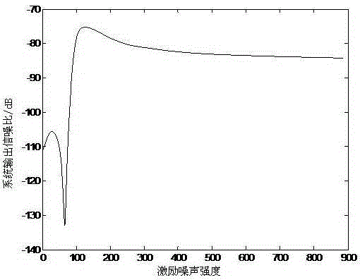
[0028] (1), establish the standard signal-to-noise ratio data collection of known lung MRI images;

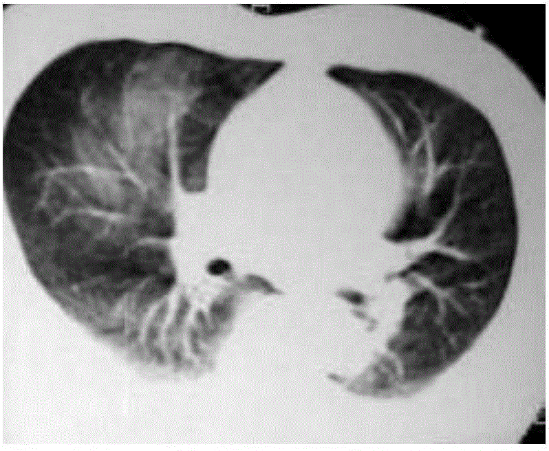
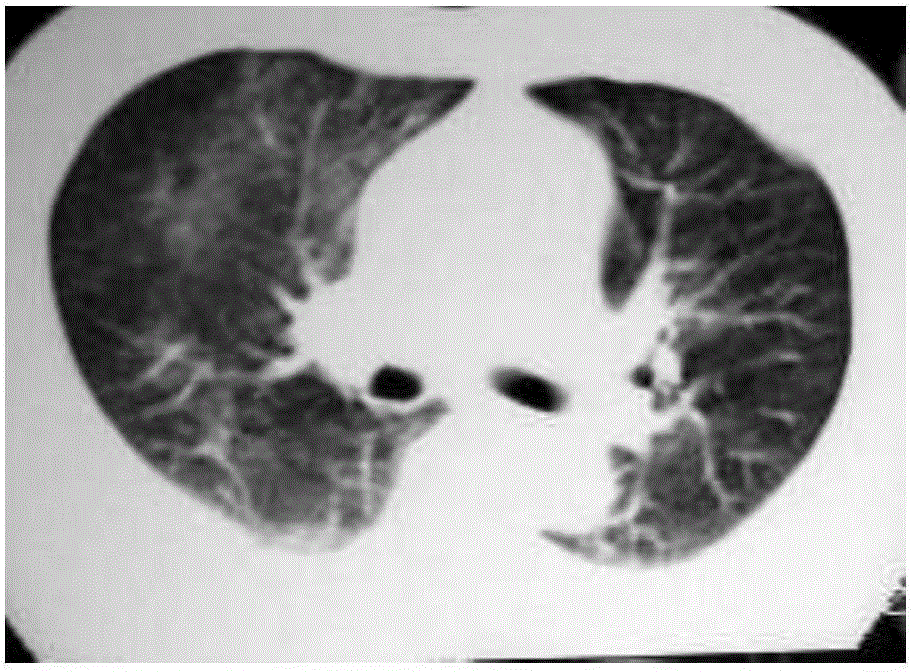
[0029] A, segment a plurality of lung MRI images by traditional method; Described traditional method is mark watershed method, also can be other conventional image segmentation methods;

[0030] B. Judging by the doctor's naked eyes whether the image segmented in step A is accurate, and if it is accurate, it will be included in the correct image set;


PUM


Abstract

The present invention relates to the technical field of image processing, and particularly to a lung tumor recognition method based on support vector machine MRI image segmentation. The method mainly adds a non-linear optimization model. A segmented image is optimized via the non-linear optimization model. The accuracy of the segmented image obtained by using this method is higher, so that the recognition accuracy of a lung tumor is higher.

Description


Claims


Application Information

Patent Timeline
Owner ZHEJIANG GONGSHANG UNIVERSITY
Why Eureka
Try Eureka