Imaging method, device and equipment and storage medium

An imaging method and image technology, applied in image enhancement, image analysis, image data processing, etc., can solve the problem that vascular map cannot assist diagnosis and treatment, and achieve the effect of facilitating diagnosis and treatment

Pending Publication Date: 2020-08-11
BEIJING NEUSOFT MEDICAL EQUIP CO LTD +1
View PDF13 Cites 0 Cited by
  • Summary
  • Abstract
  • Description
  • Claims
  • Application Information

AI Technical Summary

Benefits of technology

This technology allows for precise measurement of 3D volumes with improved accuracy compared to current methods such as X ray tomographic techniques. It involves acquiring images at multiple angles around an object while measuring its size accurately over time. By combining these measurements into one frame, it becomes possible to create virtual reality simulations that help diagnose diseases more effectively.

Problems solved by technology

This patented describes how it helps physicians make accurate medical decisions based on their understanding of blood vessel networks in order to better manage patient care more effectively. It involves studying different ways to inject fluoresce agents into arteries during surgery with an ultrasound device while watching images taken through X rays. By analyzing these images, they are able to identify areas where there may be blockages caused due to plaque buildup inside them. These blocksage locations could indicate potential issues before surgeres when trying to improve therapy efficiency.

Method used


Image

  • Imaging method, device and equipment and storage medium
  • Imaging method, device and equipment and storage medium
  • Imaging method, device and equipment and storage medium

Examples


Embodiment Construction

[0067] Reference will now be made in detail to the exemplary embodiments, examples of which are illustrated in the accompanying drawings. When the following description refers to the accompanying drawings, the same numerals in different drawings refer to the same or similar elements unless otherwise indicated. The implementations described in the following exemplary examples do not represent all implementations consistent with the present disclosure. Rather, they are merely examples of apparatuses and methods consistent with aspects of the present disclosure as recited in the appended claims.

[0068] The terminology used in the present disclosure is for the purpose of describing particular embodiments only, and is not intended to limit the present disclosure. As used in this disclosure and the appended claims, the singular forms "a", "the", and "the" are intended to include the plural forms as well, unless the context clearly dictates otherwise. It should also be understood th


PUM


Abstract

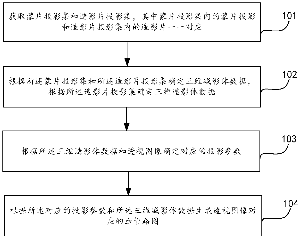
The invention provides an imaging method, device and equipment and a storage medium, and the imaging method is used for generating a blood vessel route map corresponding to a perspective image, and comprises the following steps: obtaining a mask projection set and a movie projection set, and enabling mask projections in the mask projection set to be in one-to-one correspondence with movies in themovie projection set; determining three-dimensional subtraction volume data according to the mask projection set and the radiography sheet projection set, and determining three-dimensional radiographyvolume data according to the radiography sheet projection set; determining corresponding projection parameters according to the three-dimensional radiography body data and the perspective image; andgenerating a blood vessel road map corresponding to the perspective image according to the corresponding projection parameters and the three-dimensional subtraction volume data. For perspective imagesat any angle, a blood vessel route map in the corresponding direction can be obtained, and then when the position of the rack is changed, the blood vessel route map in the corresponding position canbe correspondingly generated, and diagnosis and treatment are facilitated.

Description


Claims


Application Information

Patent Timeline
Owner BEIJING NEUSOFT MEDICAL EQUIP CO LTD
Why Eureka
Try Eureka