Ultrasound diagnostic apparatus and ultrasound image producing method

a diagnostic apparatus and ultrasound technology, applied in the field of ultrasonic diagnostic apparatus and ultrasound image producing method, can solve the problem of difficult to accurately measure the thickness of the intima-media complex of the vascular wall

Active Publication Date: 2014-03-27
FUJIFILM CORP
View PDF5 Cites 1 Cited by
  • Summary
  • Abstract
  • Description
  • Claims
  • Application Information

AI Technical Summary

Benefits of technology

The present invention provides a method for accurately determining the distance between two objects that are located within different layers of tissue or blood vessels. This helps in diagnosing diseases such as stenosis and blockages caused by plaque buildup on blood vessel walls.

Problems solved by technology

This patent describes a problem with current methods for measuring the thickness of blood vessels during medical imaging procedures. These techniques involve using ultrasounds or other types of probes to transmit ultrasonic waves towards the patient's skin and receive reflected waves from the blood vessel walls. However, these techniques often result in errors due to variations in the surrounding tissue layers caused by changes in blood flow. To address this issue, proposals include a technique called "echography," which involves analyzing the strength of sound wave signals at different locations within the blood vessel wall to estimate its thickness.

Method used


Image

  • Ultrasound diagnostic apparatus and ultrasound image producing method
  • Ultrasound diagnostic apparatus and ultrasound image producing method
  • Ultrasound diagnostic apparatus and ultrasound image producing method

Examples


Embodiment Construction

[0023]An embodiment of the present invention will now be described below based on the appended drawings.

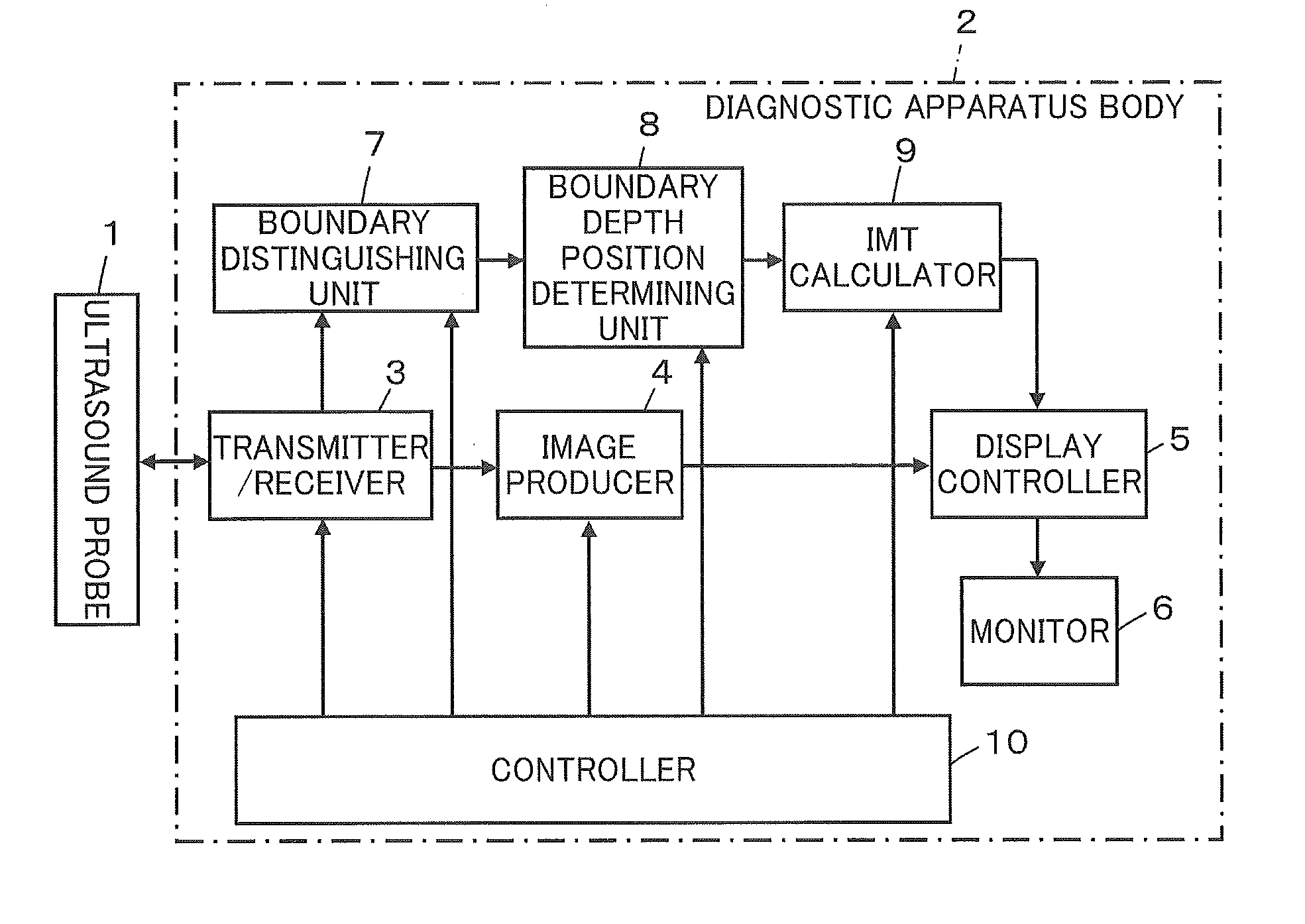
[0024]FIG. 1 shows a configuration of an ultrasound diagnostic apparatus according to the embodiment. The ultrasound diagnostic apparatus comprises an ultrasound probe 1 for transmission and reception of ultrasonic waves and a diagnostic apparatus body 2 connected to the ultrasound probe 1. The diagnostic apparatus body 2 has functions of producing an ultrasound image based on reception signals acquired by transmitting and receiving ultrasonic waves from the ultrasound probe 1 to a blood vessel in a subject and detecting boundaries of intima-media complex of a blood vessel in the ultrasound image to thereby calculate an intima-media complex thickness.

[0025]The ultrasound probe 1 is a probe of, for example, convex type, linear scan type, or sector scan type, which is brought into contact with a body surface of a subject when used. The ultrasound probe 1 comprises a plurality of ultras


PUM


Abstract

An ultrasound diagnostic apparatus includes a determining unit adapted to determine a depth position of a boundary of the intima-media complex at the clear boundary sound ray, and ascertain a depth position of the boundary of the intima-media complex at the unclear boundary sound ray; and an calculator adapted to calculate an intima-media complex thickness based on the depth positions of the boundary of the intima-media complex, wherein the determining unit performs pattern matching on a sound ray signal corresponding to the unclear boundary sound ray by using a sound ray signal corresponding to a clear boundary sound ray lying adjacent to the unclear boundary sound ray as a reference signal, thereby ascertaining the depth position of the boundary of the intima-media complex at the unclear boundary sound ray.

Description


Claims


Application Information

Patent Timeline
Owner FUJIFILM CORP
Why Eureka
Try Eureka