Femoral head lesion lesion volume measuring method based on image scanning

A measurement method and image scanning technology, applied in the field of medical image recognition, can solve problems such as cumbersome processing, large subjective interference factors, and poor human-computer interaction

Inactive Publication Date: 2021-05-28
NINGBO INST OF LIFE & HEALTH IND UNIV OF CHINESE ACAD OF SCI
View PDF0 Cites 0 Cited by
  • Summary
  • Abstract
  • Description
  • Claims
  • Application Information

AI Technical Summary

Benefits of technology

This law describes how it works on medical images that are taken from X rays or magnetic resonance imagery (MRI). By analyzing these two types of scans, we found that there were specific features within certain parts of bone structures called necks at different locations along their length. These areas could indicate diseases like osteoporosis which affect them. Based upon those findings, an equation was developed based on computed tomography (CT) scan results and machine learning techniques. It then combined multiple algorithms together to create 3 dimensional models of the entire body's bones. Using these models, researchers showed accurately where tumors had been located during knees replacement surgery. Overall, they discovered ways to improve joint health through digital protection measures such as acetabular cup prosthetic implants.

Problems solved by technology

This patented technical solution describes various techniques that aim at diagnosing and evaluating the severity of nerve damage caused by men's knee surgery during total hip replacements. These techniques include X ray fluoroscopy scans, MRI scanings, ultrasonography, computed tomographic images, etc., which provide valuable insights into cartilage degenerative processes associated with these surgeries.

Method used


Image

  • Femoral head lesion lesion volume measuring method based on image scanning
  • Femoral head lesion lesion volume measuring method based on image scanning
  • Femoral head lesion lesion volume measuring method based on image scanning

Examples


Embodiment

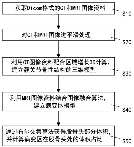
[0030] A kind of measurement method of the femoral head lesion volume based on image scanning of the present embodiment, refer to figure 1 ; including the following steps:

[0031] S10: acquiring CT and MRI image data in Dicom format;

[0032] S20: smoothing the CT and MRI images;

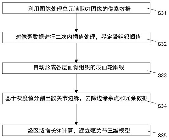
[0033] S30: Using CT image data and 3D calculation of region growth to establish a 3D model of the bony structure of the hip joint;

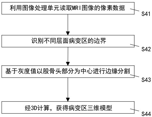
[0034] S40: Using MRI image data combined with an image fusion algorithm to establish a lesion model;

[0035] S50: Obtain the partial volume of the femoral head through the intersection algorithm of the Boolean operation, and calculate the volume ratio of the lesion area at the femoral head.

[0036] Among them, after the 3D model of the hip joint is established, when the 3D model of the femoral head is separated and obtained by the intersection algorithm of Boolean operations, the mask of the proximal femur is first separated based on the gray value segmentation, a


PUM


Abstract

The invention relates to the technical field of medical image recognition, and provides a femoral head lesion volume measurement method based on image scanning, which comprises the following operation steps: acquiring CT and MRI image data in a Dicom format; carrying out smoothing processing on the CT and MRI images; establishing a three-dimensional model of a hip joint bony structure by utilizing CT image data in cooperation with region growing 3D calculation; establishing a lesion area model by using MRI image data in combination with an image fusion algorithm; and obtaining the volume of the femoral head part through an intersection algorithm of Boolean operation, and calculating the volume proportion of the lesion area at the femoral head. The objective of the invention is to provide a scientific measurement method for realizing positioning and size visualization of a lesion area, and more accurate data is provided for formulating a hip protection treatment scheme.

Description


Claims


Application Information

Patent Timeline
Owner NINGBO INST OF LIFE & HEALTH IND UNIV OF CHINESE ACAD OF SCI
Why Eureka
Try Eureka