Thoracolumbar vertebra trauma CT image evaluation method and system
A CT imaging and evaluation system technology, applied in the field of medical information, can solve the problems such as the inability to automatically generate TLICS qualitative evaluation, and the inability to automatically detect important dangerous signs.
- Summary
- Abstract
- Description
- Claims
- Application Information
AI Technical Summary
Problems solved by technology
Method used
Image

Examples
Embodiment 1
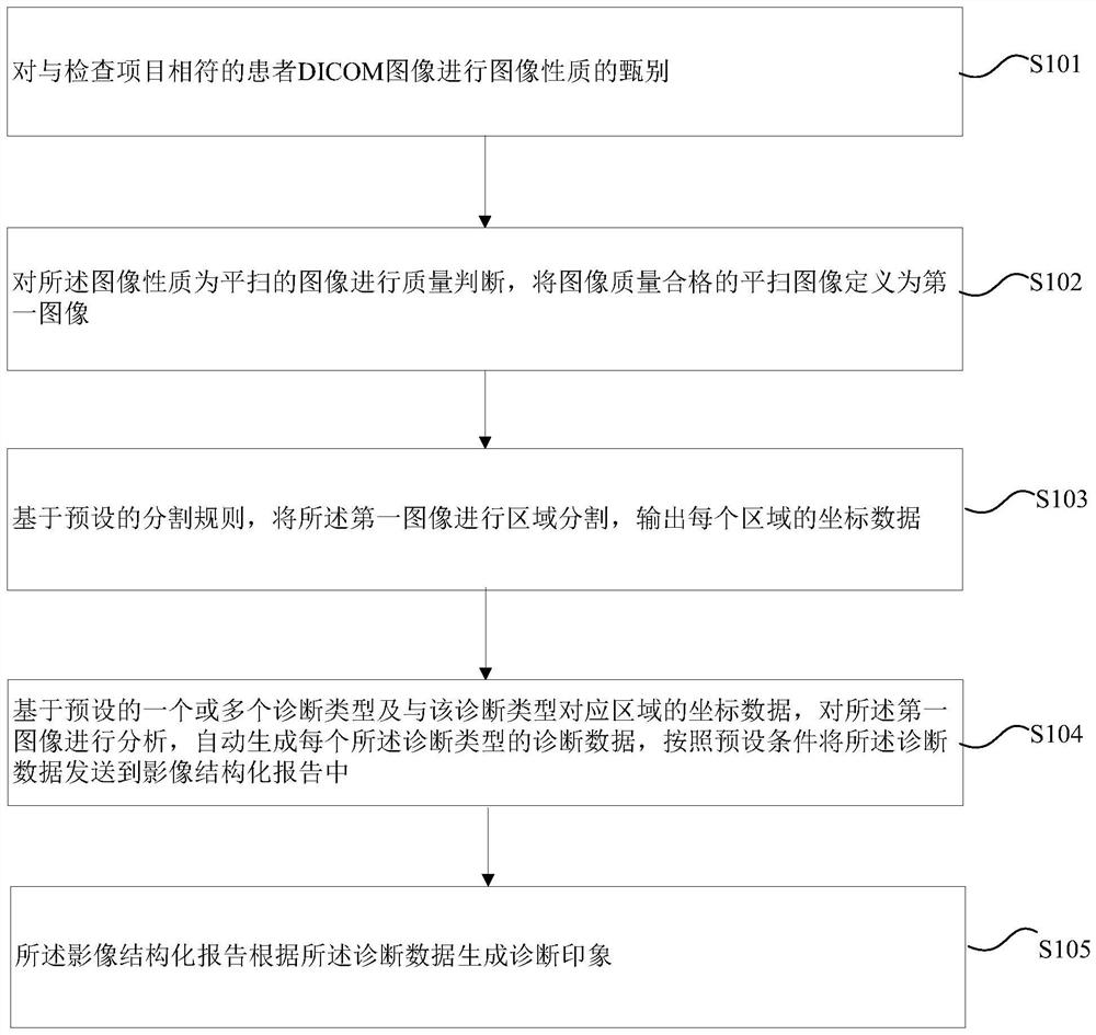
[0025] figure 1 It shows a flow chart of the CT image evaluation method for thoracolumbar trauma according to Embodiment 1 of the present invention; figure 1 As shown, the method includes the following steps:
[0026] Step S101, discriminating the image properties of the patient's DICOM images that match the inspection items;
[0027] Among them, the DICOM image of the patient that matches the inspection items is used to judge whether it matches the inspection items registered in the RIS. The tools used in this process are program and AI model recognition. Input patient DICOM images, output grouped images, qualitative judgment-check items, prompt information-check items.
[0028] Qualitative judgments are returned to the corresponding controls of the structured report "Technical Assessment".
[0029] If it is judged that the DICOM image of the patient is consistent with the inspection items, the output image will be used in the subsequent AI diagnosis process.
[0030] If it
Embodiment 2
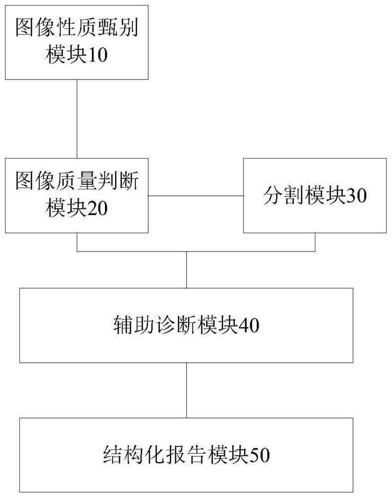
[0078] figure 2 It shows a schematic structural diagram of a CT image evaluation system for thoracolumbar trauma according to Embodiment 2 of the present invention; figure 2 As shown, the system includes: an image property screening module 10, an image quality judgment module 20, a segmentation module 30, an auxiliary diagnosis module 40 and a structured report module 50, wherein,
[0079] The image property discrimination module 10 is connected to the image quality judgment module 20, and is used to discriminate the image property of the patient's DICOM images that match the inspection items;
[0080] Among them, the DICOM image of the patient that matches the inspection items is used to judge whether it matches the inspection items registered in the RIS. The tools used in this process are program and AI model recognition. Input patient DICOM images, output grouped images, qualitative judgment-check items, prompt information-check items.
[0081] Qualitative judgments are r
Embodiment 3
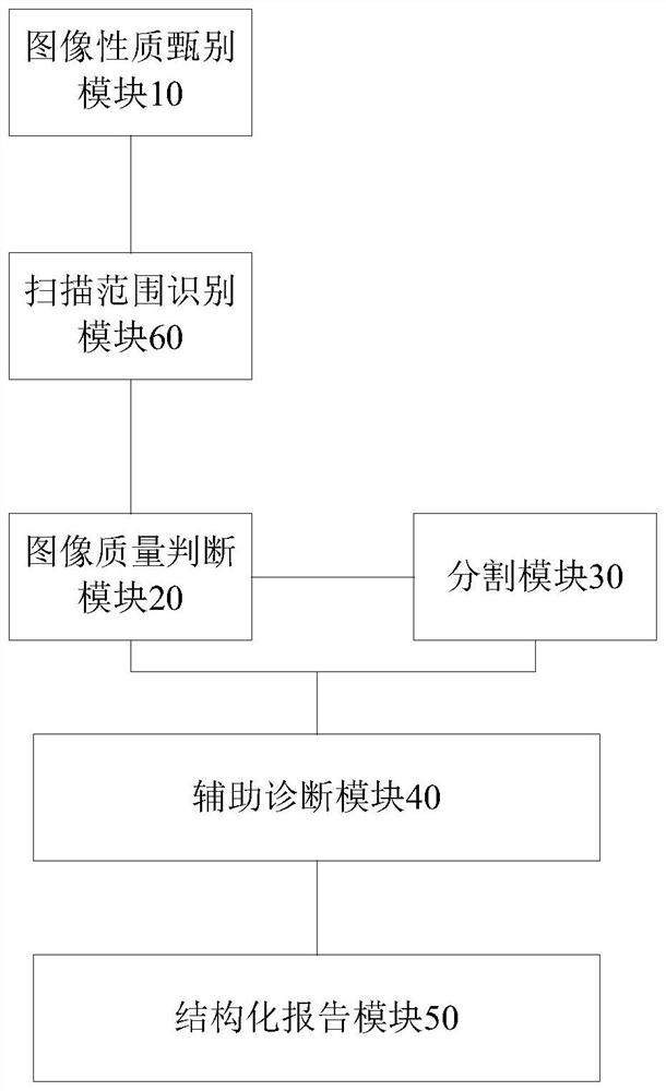
[0130] image 3 It shows a schematic structural diagram of a CT image evaluation system for thoracolumbar trauma according to Embodiment 3 of the present invention; image 3 As shown, the system also includes: a scan range identification module 60, which is connected to the image quality judgment module 20 and the segmentation module 30 respectively, and is used to identify The scan range of the DICOM image of the patient, wherein the scan range is: thoracic spine, thoracolumbar segment, lumbar spine, and lumbosacral spine.
[0131] The tool used in this step is AI model recognition, input plain scan image, output qualitative judgment-scan range, images of different scan ranges are used for subsequent segmentation models, and output different positioning values.
[0132] From the above description, it can be seen that the above-mentioned embodiments of the present invention have achieved the following technical effects: the embodiments of the present invention apply AI models, r
PUM
 Abstract
Description
Claims
Application Information
 - R&D Engineer
- R&D Manager
- IP Professional
- Industry Leading Data Capabilities
- Powerful AI technology
- Patent DNA Extraction
Browse by: Latest US Patents, China's latest patents, Technical Efficacy Thesaurus, Application Domain, Technology Topic.
© 2024 PatSnap. All rights reserved.Legal|Privacy policy|Modern Slavery Act Transparency Statement|Sitemap