Ultrasound diagnostic apparatus and method of controlling ultrasound diagnostic apparatus
a diagnostic apparatus and ultrasound technology, applied in diagnostics, medical science, applications, etc., can solve the problems of inability to obtain appropriate test results, less skilled users may perform compression tests without moving the position of ultrasound probes from the back of the knee,
- Summary
- Abstract
- Description
- Claims
- Application Information
AI Technical Summary
Benefits of technology
Problems solved by technology
Method used
Image

Examples
embodiment 1
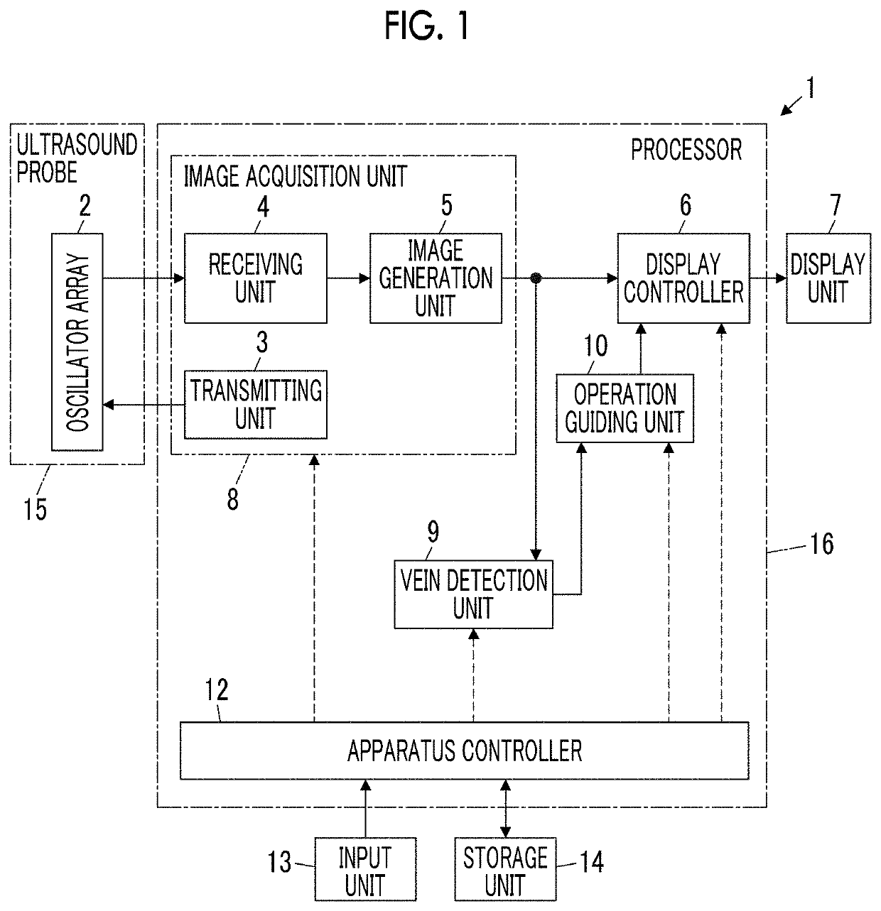
[0029]A configuration of an ultrasound diagnostic apparatus 1 according to Embodiment 1 of the present invention is shown in FIG. 1. As shown in FIG. 1, the ultrasound diagnostic apparatus 1 comprises an oscillator array 2, and a transmitting unit 3 and a receiving unit 4 are connected to the oscillator array 2. An image generation unit 5, a display controller 6, and a display unit 7 are sequentially connected to the receiving unit 4. An image acquisition unit 8 is configured by the transmitting unit 3, the receiving unit 4, and the image generation unit 5. A vein detection unit 9 is connected to the image generation unit 5, and an operation guiding unit 10 is connected to the vein detection unit 9. The operation guiding unit 10 is connected to the display controller 6.
[0030]The apparatus controller 12 is connected to the display controller 6, the image acquisition unit 8, the vein detection unit 9, the operation guiding unit 10, and the input unit 13 and the storage unit 14 are connec
embodiment 2
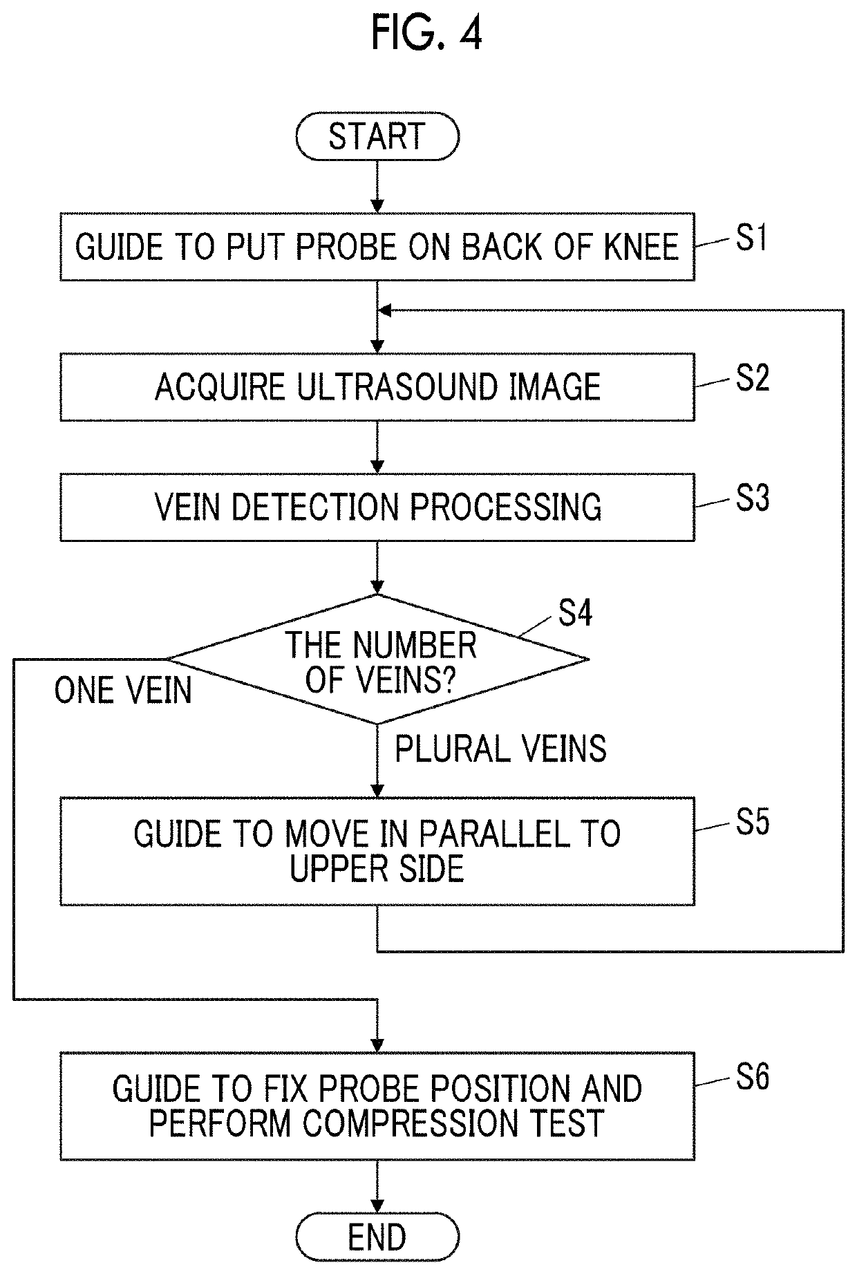
[0072]In Embodiment 1, in a case where a plurality of popliteal veins are detected by the vein detection unit 9, until only one popliteal vein is detected, the ultrasound image is acquired while moving the ultrasound probe 15 to an upper side of the back of the knee, but the movement range of the ultrasound probe 15 can be determined such that the ultrasound probe 15 does not move too much upward.
[0073]The configuration of an ultrasound diagnostic apparatus 1A according to Embodiment 2 is shown in FIG. 7. The ultrasound diagnostic apparatus 1A of Embodiment 2 comprises an apparatus controller 12A instead of the apparatus controller 12 in the ultrasound diagnostic apparatus 1 of Embodiment 1 shown in FIG. 1, and further comprises a probe movement range determining unit 22.
[0074]In the ultrasound diagnostic apparatus 1A according to Embodiment 2, the probe movement range determining unit 22 is connected to the operation guiding unit 10. The apparatus controller 12A is connected to the di
PUM
 Abstract
Description
Claims
Application Information
 - R&D Engineer
- R&D Manager
- IP Professional
- Industry Leading Data Capabilities
- Powerful AI technology
- Patent DNA Extraction
Browse by: Latest US Patents, China's latest patents, Technical Efficacy Thesaurus, Application Domain, Technology Topic.
© 2024 PatSnap. All rights reserved.Legal|Privacy policy|Modern Slavery Act Transparency Statement|Sitemap