Promotion of brain self-repair mechanisms by stereotaxic micro-stimulation

a stereotaxic micro-stimulation and brain technology, applied in the field of brain self-repair mechanisms, can solve the problems of impaired memory, thinking and behavior, impaired brain synaptic activity, and stiff joints

Inactive Publication Date: 2013-05-28
UNIV OF SOUTH FLORIDA +1
View PDF8 Cites 1 Cited by
  • Summary
  • Abstract
  • Description
  • Claims
  • Application Information

AI Technical Summary

Benefits of technology

This patent describes how tiny needles can be used to help regulate nerves in the brain. They have small needles with very thin needles made up of materials like gold or platinum. These needles are placed slowly over time and moved around inside the brain where neurons were located. By inserting them into specific areas of the brain, these needles could cause new connections between different parts of the body.

Problems solved by technology

This patent describes methods for promoting differentiation of stem cells called BSCs, which play key roles in maintaining homeostasis within the body during development and regeneration processes. The technical problem addressed in this patent is how to efficiently induce endogenous stem cells to differentiate into neural precursors, thereby promoting the self-repair mechanisms of the brain.

Method used


Image

  • Promotion of brain self-repair mechanisms by stereotaxic micro-stimulation
  • Promotion of brain self-repair mechanisms by stereotaxic micro-stimulation
  • Promotion of brain self-repair mechanisms by stereotaxic micro-stimulation

Examples


example

Mobilization of GFP+ Cells from the Periphery in to the CNS Following Focal Micro-Lesions of Brain

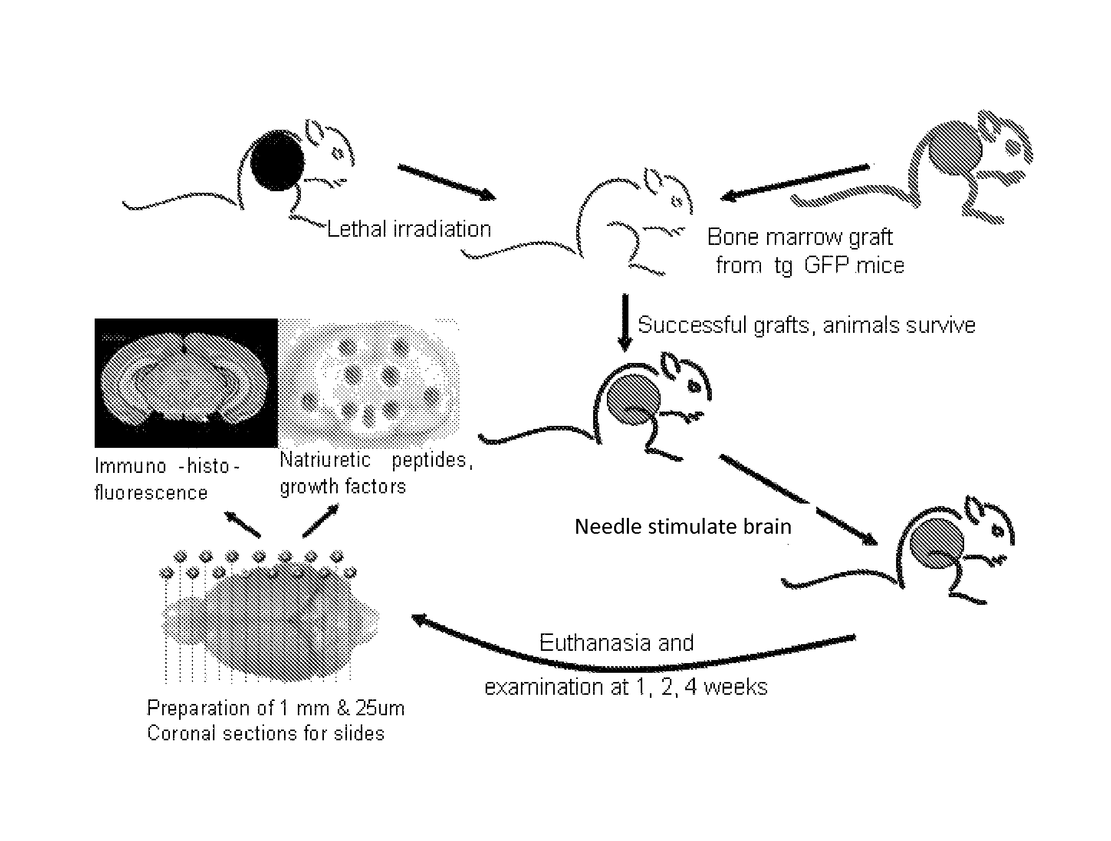
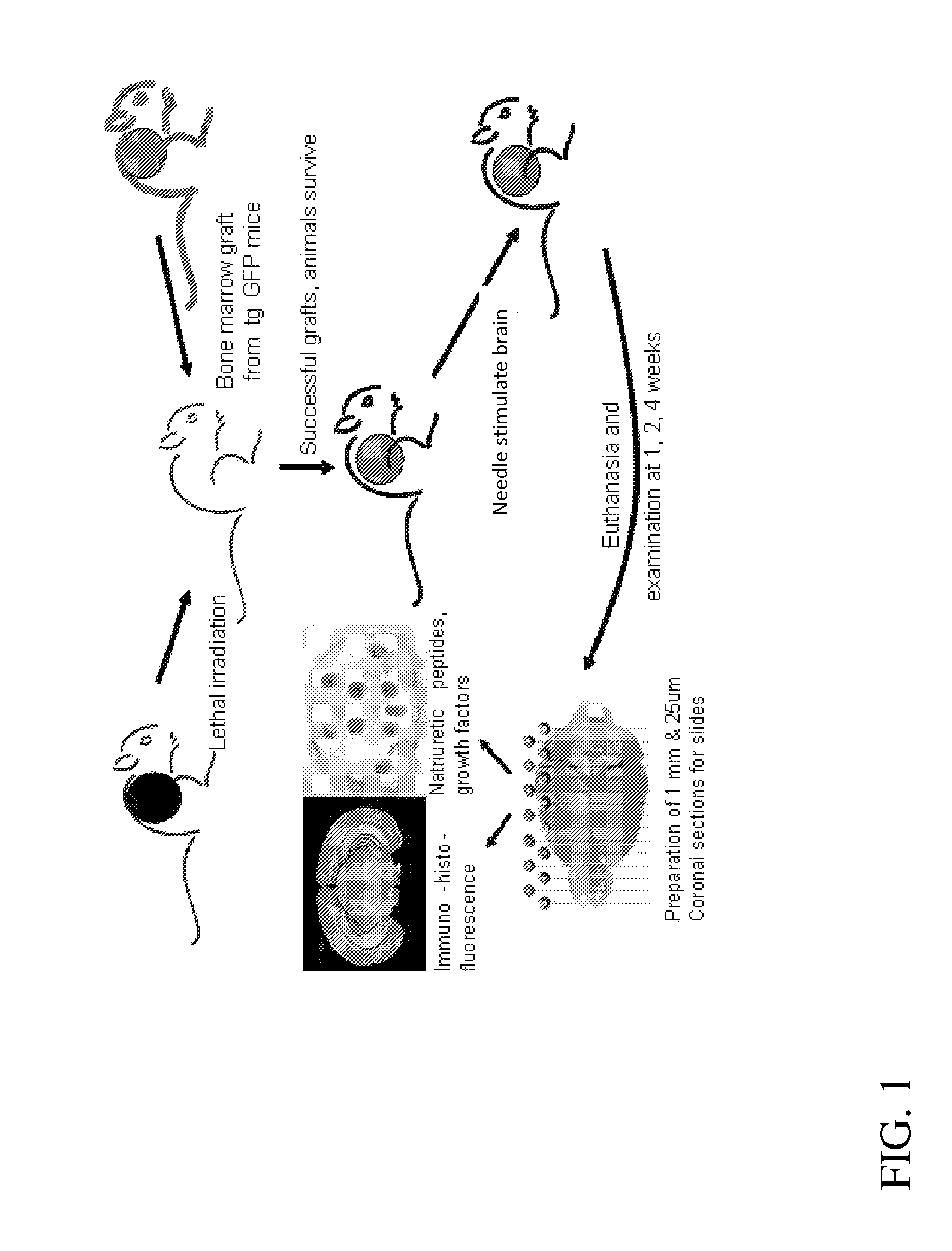
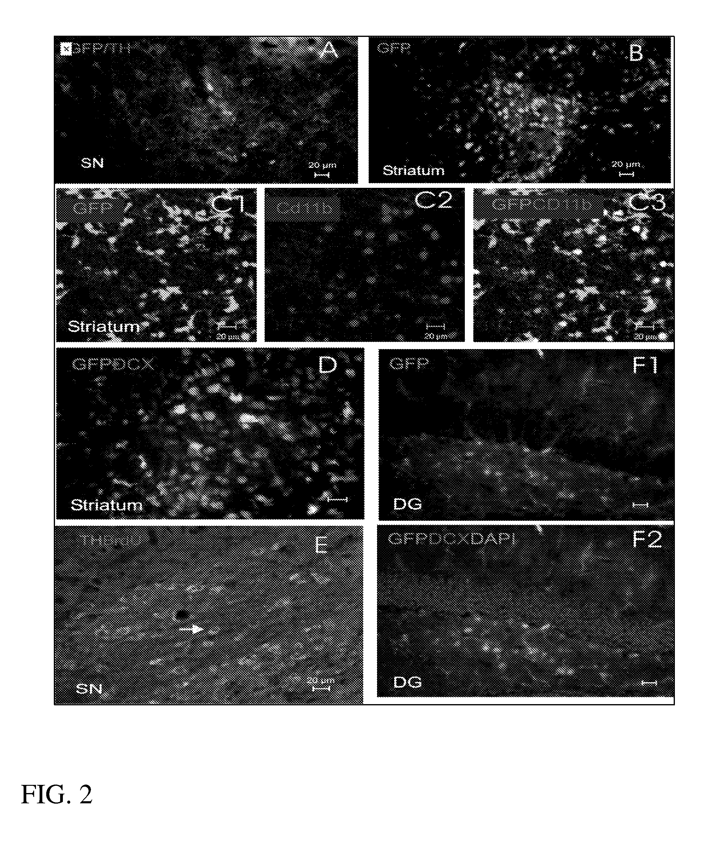
[0073]A sterile micro-needle (200 microns maximum shaft diameter, similar to the acupuncture needle) was transiently inserted stereotaxically into four different sites: corpus striatum, dorsal hippocampus, cerebellum and ventral midbrain. Several of the mice underwent bromodeoxyuridine (BrdU) injections (100 mg / kg i.p.×2) on the day of transient insertion of the needle to label newly born cells. One, two and four weeks after making the micro-lesions, mice were euthanatized and their brains were processed for immunohistochemistry. GFP+ cells were readily visualized at the site of the micro-lesions in striatum, ventral midbrain and hippocampus (FIG. 2). The GFP+ cells could be found along the tract of the needle and extending beyond the locus of the lesion. In the striatal lesion (FIG. 2B, 2C) many of the GFP+ cells that had infiltrated the lesion from the peripheral circulation bore the m


PUM


Abstract

A method of treating neurological deficit in which the brain area affected is focally and precisely stimulated by the transient insertion and subsequent removal of a micro-needle. This insertion and subsequent removal of the micro-needle induces endogenous stem cells to proliferate, migrate and promote the brain's self-repair mechanisms. The micro-needle stimulation causes the birth of new neural cells within the brain as well as mobilizes bone marrow derived cells with a neuronal phenotype to migrate to the site of stimulation to repair and replace damaged neural cells. By repairing and/or replacing injured or dead cells, this approach will slow down the degenerative course of the disease and may result in reversal of symptoms.

Description


Claims


Application Information

Patent Timeline
Owner UNIV OF SOUTH FLORIDA
Why Eureka
Try Eureka