X-ray imaging system with optical auxiliary calibration

An optical imaging and imaging technology, applied in the field of X-ray imaging, can solve the problems of harming the body, large radiation dose, and increasing the cost of radiology rooms, and achieve the effect of protecting doctors and high universality

Inactive Publication Date: 2014-11-19
SHENZHEN INST OF ADVANCED TECH CHINESE ACAD OF SCI
View PDF4 Cites 3 Cited by
  • Summary
  • Abstract
  • Description
  • Claims
  • Application Information

AI Technical Summary

Benefits of technology

This new technology allows for better monitoring during medical procedures by combining two different types of cameras together into one device that provides real time image data without exposing them physically or electronics near their location.

Problems solved by technology

This patents describes how current x- ray imaging facilities lack an individual patient's location monitoring function due to their high costs associated with opening windows or exposing patients during operation.

Method used


Image

  • X-ray imaging system with optical auxiliary calibration
  • X-ray imaging system with optical auxiliary calibration

Examples


Embodiment Construction

[0024] Hereinafter, embodiments of the present invention will be described in detail with reference to the accompanying drawings.

[0025] The difference between X-ray and visible light lies in the different wavelengths. It does not deflect in the electric field or magnetic field, and can reflect, refract, interfere, diffract, etc.; it has the ability to penetrate substances, but its penetration ability is different for different substances. . Therefore, X-rays can make the human body form an image on the screen or film, which is based on the difference in density and thickness of human tissue.

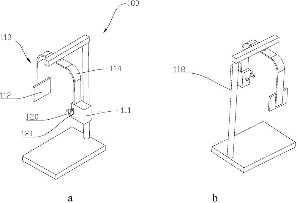
[0026] In a common X-ray system, an operator (such as a radiologist) usually directs an X-ray source to an imaging object (such as a part of a patient to be imaged) visually, and then performs imaging. In this case, the operator must be able to directly observe the imaging situation at the shooting site, and sometimes even need to take a test shot to confirm the accuracy of the position


PUM


Abstract

The invention relates to the field of X-ray imaging technologies, in particular to an X-ray imaging system with the optical auxiliary calibration. The system comprises an X-ray imaging module, a visible ray imaging device and a remote processing terminal. The X-ray imaging module comprises an X-ray source and an X-ray imaging device oppositely corresponding to the X-ray source and is used for collecting an X-ray image of an imaged object placed between the X-ray source and the X-ray imaging device. The visible ray imaging device and the X-ray source are arranged in the same direction, and the visible ray imaging device is used for collecting an image of the imaged object at the same body position. The remote processing terminal is respectively connected with the X-ray imaging module and the visible ray imaging device and used for performing relative bias difference value calculation on the received X-ray image and the image so that the X-ray image and the image can coincide after adjusting the relative bias difference value. From the point of view of safety and practicability, it can be guaranteed that a doctor effectively obtains the pose of a patient in real time, the structure of a radiation room can be simplified, and besides the safety of the doctor can be improved.

Description


Claims


Application Information

Patent Timeline
Owner SHENZHEN INST OF ADVANCED TECH CHINESE ACAD OF SCI
Why Eureka
Try Eureka