Auxiliary device for minimally invasive surgery

An auxiliary device and minimally invasive surgery technology, applied in the field of medical devices, can solve the problems of inconvenience for doctors to cut biological tissue, difficult to distinguish the boundary of diseased tissue, inaccurate cutting of diseased tissue, etc., so as to facilitate the operation of doctors, improve magnetic force and grasp strength, the effect of improving the success rate

Pending Publication Date: 2020-03-24
SHENZHEN JIFU MEDICAL TECH CO LTD
View PDF0 Cites 5 Cited by
  • Summary
  • Abstract
  • Description
  • Claims
  • Application Information

AI Technical Summary

Benefits of technology

This patented technology helps surgeons perform minimal-invasively procedures more efficiently while also improving their ability to control electrical shocks during medical interventions. It uses an adapter that attaches two smaller permanent magnets together through strong magnesis or other means like attraction between them. When there are no enough powerful magents available at all times, another larger one will deliver it instead.

Problems solved by technology

Technicians face challenges with finding ways to securely hold biological samples during procedures such as gastronerosomy surgeries because current devices require manual laborers' manipulation over many hours per day. Additionally, existing methods involve attaching permanent magnebiles onto metal clasches made from nonmetallizable materials like stainless steel, making them hard to clean after each procedure. There needs a better way to attach these temporary objects without requiring excessive effort and reducing operational efficacy compared to traditional solutions involving separate tools.

Method used


Image

  • Auxiliary device for minimally invasive surgery
  • Auxiliary device for minimally invasive surgery
  • Auxiliary device for minimally invasive surgery

Examples


Embodiment Construction

[0044] In order to make the technical problems, technical solutions and beneficial effects to be solved by the present invention clearer, the present invention will be further described in detail below in conjunction with the accompanying drawings and embodiments. It should be understood that the specific embodiments described here are only used to explain the present invention, not to limit the present invention.

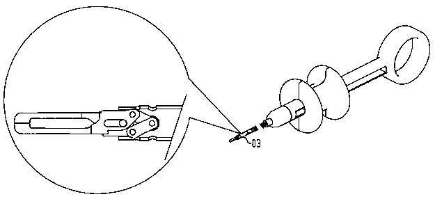
[0045] Please refer to Figure 1 to Figure 3 , are respectively the schematic diagram of the initial state, the schematic diagram of the closed state and the schematic diagram of the locked state of the auxiliary device for minimally invasive surgery of the present invention, Figure 4 It is a sectional view of the front end of the clip of the auxiliary device for minimally invasive surgery of the present invention, and further reference Figure 5 Schematic diagram of the clamping part of the auxiliary device. The auxiliary device for minimally invasive surgery inclu


PUM


Abstract

The invention discloses an auxiliary device for minimally invasive surgery. The device comprises a clamping part, a locking part, a control line, a bendable catheter and a control part. A small magnetcan be further arranged at the rear end of the locking part. The control line controls the clamping part to open and close by taking gaps formed between two sides of the small magnet and the inner wall of the locking part as channels. An open circular groove in the front end of the control line is connected with a rear-end pin. The clamping part is driven by a connecting rod. When the control line moves towards the rear end, the clamping part is locked, and the connecting rod protrusion is clamped into a locking groove of a clamp cylinder. The control line is separated from the rear-end pin,and finally the control line presses an elastic buckle at the front end of a spring line. The bendable catheter is separated from the clamp cylinder. A standby magnet can be further added to the auxiliary device to improve magnetic force. The auxiliary device for minimally invasive surgery can accurately anchor and lift biological tissues, and is convenient for doctors to operate. The surgery efficiency and success rate are improved.

Description


Claims


Application Information

Patent Timeline
Owner SHENZHEN JIFU MEDICAL TECH CO LTD
Why Eureka
Try Eureka