Preparation method of mouse bone marrow macrophages

A technique for macrophages and mice, applied in the field of preparation of mouse bone marrow macrophages

Pending Publication Date: 2020-05-22
NINGXIA UNIVERSITY
View PDF2 Cites 0 Cited by
  • Summary
  • Abstract
  • Description
  • Claims
  • Application Information

AI Technical Summary

Benefits of technology

This technology allows scientists to quickly separate mice from their own blood cells that are used during research or testing them on other animals more easily than before they were infected with viruses like measles virus (measuring how well people have been immune).

Problems solved by technology

The technical problem addressed in this patented text relates to developing efficient ways to isolate specific types of primordially dividing mononuclear giant leukemia stem cells called TSCBMCs, particularly those containing certain genes associated with metabolism. This requires identifying these specialized subsets of primitive megakoblasts during early stages of embryonic ontogeneesis process. Current methodologies involve culturing isolated rat hepatoma-like medullum projections onto plastic surfaces like glass slides, flasks, and tubular membranes, but there currently exist challenges involving handling and manipulation steps required beforehand.

Method used


Image

  • Preparation method of mouse bone marrow macrophages
  • Preparation method of mouse bone marrow macrophages

Examples


preparation example Construction

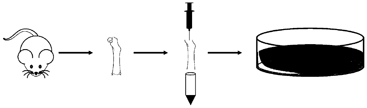
[0023] The invention provides a method for preparing mouse bone marrow macrophages, comprising the following steps:

[0024] (1) preparing a DMEM medium containing 10% fetal bovine serum to obtain a complete medium, and putting the mouse fibroblast L929 cell culture supernatant and the complete medium into a water bath for incubation;

[0025] (2) Mice 6-9 weeks old were killed by cervical dislocation; soaked in 75% ethanol for 2-3 minutes;

[0026] (3) Sterilized surgical scissors to peel off the skin and muscles of the mouse legs, cut the femur and tibia of the mouse, scrape off the muscle and fascia on the bone as much as possible, and use phosphate buffer containing 2% penicillin and streptomycin Fluid rinses the bone surface;

[0027] (4) Cut off both ends of the femur and tibia with surgical scissors; absorb DMEM medium with a 5mL disposable syringe, insert the needle of the syringe into the medullary cavity of the femur and tibia accurately, blow out the bone marrow, repe

Embodiment

[0035] (1) preparing a DMEM medium containing 10% fetal bovine serum to obtain a complete medium, and putting the mouse fibroblast L929 cell culture supernatant and the complete medium into a water bath for incubation;

[0036] (2) Mice 6-9 weeks old were killed by cervical dislocation; soaked in 75% ethanol for 2-3 minutes;

[0037] (3) Sterilized surgical scissors to peel off the skin and muscles of the mouse legs, cut the femur and tibia of the mouse, scrape off the muscle and fascia on the bone as much as possible, and use phosphate buffer containing 2% penicillin and streptomycin Fluid rinses the bone surface;

[0038] (4) Cut off both ends of the femur and tibia with surgical scissors; absorb DMEM medium with a 5mL disposable syringe, insert the needle of the syringe into the medullary cavity of the femur and tibia accurately, blow out the bone marrow, repeat several times until the femur and tibia are colorless, and collect after blowing Transfer the single cell suspensio


PUM


Abstract

The invention provides a preparation method of mouse bone marrow macrophages. The preparation method comprises the steps as follows: (1) preparing a complete medium; (2) dislocating cervical vertebraof mice of 6-9 weeks old; (3) taking femur and tibia of the mice, blowing out mouse bone marrow cells, repeating the operation until the bone is colorless, collecting a single cell suspension after blowing, performing centrifuging, and discarding a supernatant; (4) adding a red blood cell lysis buffer to cell precipitate for treatment; (5) adding the mixed solution to a complete medium rapidly forneutralization, performing centrifuging, and discarding a supernatant; (6) adding the complete medium to centrifugal cell precipitate to resuspend cells; (7) inoculating a petri dish with the resuspended cells for culture; (8) after 4 hours, sucking out the suspension cells, inoculating a petri dish of the complete medium containing an inducer with the cells, and recording the day as the first day; (9) changing half of the medium of the cells on the third day, changing the medium every other day until the seventh day, and performing testing. A simple, economical and rapid method for separating the mouse bone marrow macrophages is provided.

Description


Claims


Application Information

Patent Timeline
Owner NINGXIA UNIVERSITY
Why Eureka
Try Eureka