Three-dimensional medical image mark point extraction method and system

A medical image and three-dimensional image technology, applied in the field of medical image processing, can solve problems such as uneven reconstruction artifacts, and achieve the effects of improving extraction accuracy, high operating efficiency, and improving system accuracy.

Active Publication Date: 2021-07-13
SHANGHAI DROIDSURG MEDICAL CO LTD
View PDF10 Cites 3 Cited by
  • Summary
  • Abstract
  • Description
  • Claims
  • Application Information

AI Technical Summary

Benefits of technology

This patented technology allows for accurate detection of markers points that have been marked onto an object during surgery without having any complicated operations involved. It achieves this by scanning multiple images taken at different angles around the patient's body while capturing them continuously over time. These images can then be analyzed together to determine if they match up correctly. Overall, it improves the precision and reliability of detecting these marks accurately throughout surgeries.

Problems solved by technology

Technics: Current techniques involve markers that help guide surgeries accurately during procedures like endoscopy (endoscopic examination). These markers include gold balls placed at specific locations along certain lines within the body where they were inserted into tubular organs such as blood vessels or lungs. They also work well when attached correctly but may cause issues if improperly aligned because these marks cannot easily distinguish them from surrounding tissue structures.

Method used


Image

  • Three-dimensional medical image mark point extraction method and system
  • Three-dimensional medical image mark point extraction method and system
  • Three-dimensional medical image mark point extraction method and system

Examples


no. 1 example

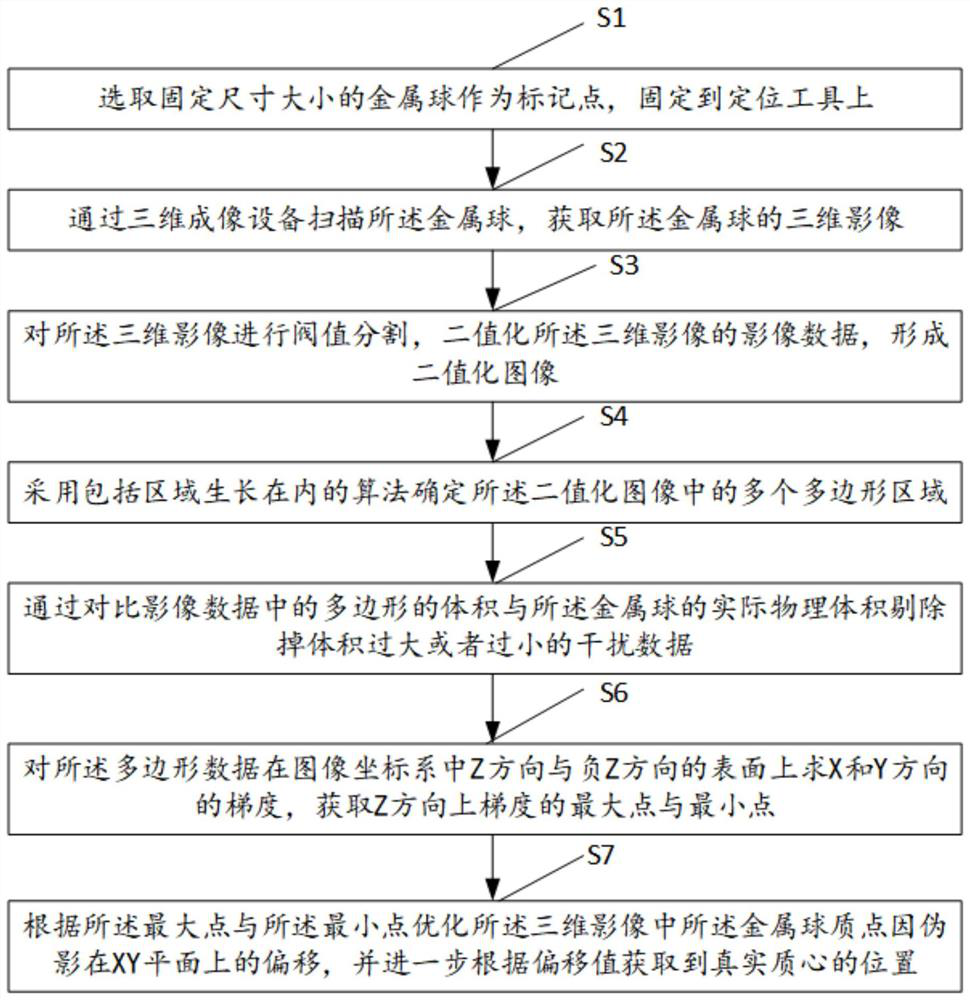
[0056] Such as figure 1 As shown, the present embodiment provides a method for extracting marker points from three-dimensional medical images, comprising the following steps:

[0057] S1: Select a metal ball with a fixed size as a marking point and fix it on the positioning tool.

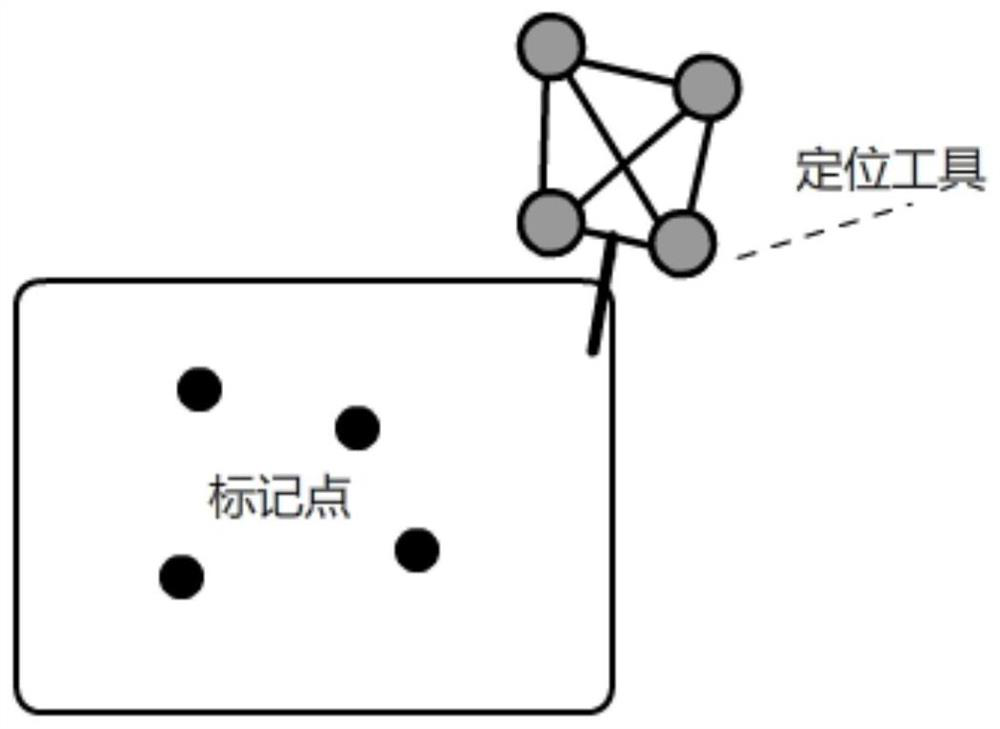
[0058] Specifically, in this embodiment, as figure 2 As shown, generally, the metal ball that is most easily recognized by the 3D imaging device is selected as the marking point of the positioning tool. The metal ball is fixed on the positioning tool, and the positioning tool is fixed on the object to be calibrated. After the position of the metal ball is recognized by the three-dimensional imaging device, the coordinates of the object to be calibrated can be obtained through the coordinate transformation relationship between the metal ball and the positioning tool, and the coordinate transformation relationship between the positioning tool and the object to be calibrated.

[0059] When obtaining th


PUM


Abstract

The invention relates to the technical field of medical image processing, and provides a three-dimensional medical image mark point extraction method and system, and the method comprises the steps of scanning a metal ball through a three-dimensional imaging device, and obtaining a three-dimensional image of the metal ball; performing threshold segmentation on the three-dimensional image, and binarizing image data of the three-dimensional image to form a binarized image; determining a plurality of polygonal regions in the binarized image; comparing the volume of the polygon in the image data with the actual physical volume of the metal ball, and eliminating interference data with too large or too small volume; solving gradients of the polygon data in the X direction and the Y direction on the surfaces of the Z direction and the negative Z direction in the image coordinate system, and obtaining a maximum point and a minimum point of the gradients in the Z direction; and according to the maximum point and the minimum point, optimizing the offset of the metal sphere mass point in the three-dimensional image on the XY plane due to the artifact, and further obtaining the position of the true centroid according to the offset value. A mass point extraction method is optimized to a certain extent, and the extraction precision of the mark points is improved.

Description


Claims


Application Information

Patent Timeline
Owner SHANGHAI DROIDSURG MEDICAL CO LTD
Why Eureka
Try Eureka