Kit for detecting hepatitis B virus and detection method of hepatitis B virus
A hepatitis B virus and kit technology, applied in biochemical equipment and methods, microbial determination/inspection, etc., can solve the problems of difficulty in detection, false positives, and time-consuming, and achieve improved detection sensitivity, low cost, and rapid detection. Effect
- Summary
- Abstract
- Description
- Claims
- Application Information
AI Technical Summary
Problems solved by technology
Method used
Image

Examples
preparation example Construction
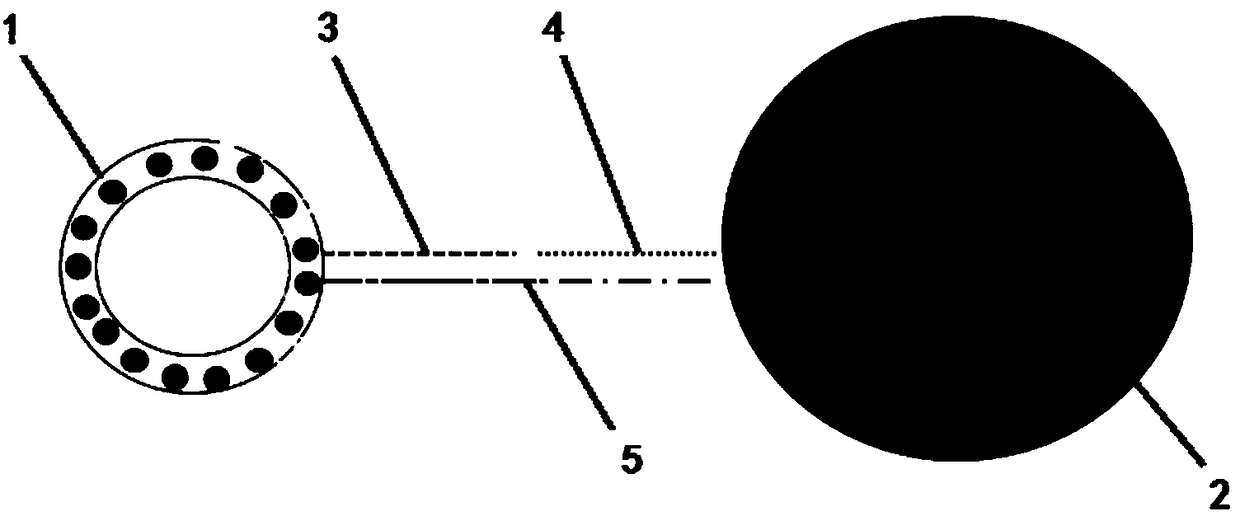
[0029] In the present invention, the liposome-QD complex is carboxyl functionalized liposome quantum dot microspheres. The preparation method of the liposome-QD complex comprises: mixing distearoyl phosphatidylcholine, phospholipid-polyethylene glycol-carboxyl, cholesterol and CdSe quantum dots in chloroform, and preparing lipid by film hydration method. Plastid-QD complexes. Described distearoyl phosphatidylcholine, phospholipid-polyethylene glycol-carboxyl and cholesterol are the membrane material of making liposome, described distearoyl phosphatidylcholine, phospholipid-polyethylene glycol-carboxyl, The molar ratio of cholesterol to the quantum dots is 9-12:2-4:0.8-1.5:4-6.5, preferably 10-11:2.5-3.5:0.9-1.2:4.5-6. The mixing temperature is preferably 35-45°C, more preferably 38-42°C. In the present invention, the emission wavelength of the CdSe quantum dots is 488nm, 561nm or 647nm. Different emission wavelengths can be excited with the same excitation light, observed in d
Embodiment 1
[0045] A test kit for detecting hepatitis B virus, comprising: a liposome-QD complex-labeled reporter probe, a magnetic bead-modified capture probe and a buffer; the reporter probe nucleic acid sequence and the capture probe nucleic acid sequence Complementary to HBV DNA respectively. Its preparation method is as follows:
[0046] 1. Preparation of liposomal quantum dot microspheres:
[0047] Add 5mL chloroform to the long-necked flask, then add 0.1M distearoyl phosphatidylcholine 100μl, 0.1M phospholipid-polyethylene glycol-carboxyl 36ul, 50mM cholesterol 22ul, 58ul, 0.1M CdSe quantum dots with an emission wavelength of 488nm, Mix well at a temperature of 40°C. The chloroform was evaporated under reduced pressure with a rotary evaporator, and when a film appeared on the inner wall of the long-necked flask, it was dried with argon. Then add 2 mL of LPBS (pH7.4), shake and dissolve the film at 30°C, and filter the dissolved solution three times with a 0.2 μm polycarbonate membr
Embodiment 2
[0055] A test kit for detecting hepatitis B virus, comprising: a liposome-QD complex-labeled reporter probe, a magnetic bead-modified capture probe and a buffer; the reporter probe nucleic acid sequence and the capture probe nucleic acid sequence Complementary to HBV DNA respectively. Its preparation method is as follows:
[0056] 1. Preparation of liposomal quantum dot microspheres:
[0057] Add 5mL chloroform to the long-necked flask, then add 0.1M distearoylphosphatidylcholine 100μl, 0.1M phospholipid-polyethylene glycol-carboxylate 36ul, 50mM cholesterol 22ul, 58ul, 0.1M CdSe quantum dots with an emission wavelength of 561nm, Mix well at a temperature of 40°C. The chloroform was evaporated under reduced pressure with a rotary evaporator, and when a film appeared on the inner wall of the long-necked flask, it was dried with argon. Then add 2 mL of LPBS (pH7.4), shake and dissolve the film at 30°C, and filter the dissolved solution three times with a 0.2 μm polycarbonate mem
PUM
 Abstract
Description
Claims
Application Information
 - R&D Engineer
- R&D Manager
- IP Professional
- Industry Leading Data Capabilities
- Powerful AI technology
- Patent DNA Extraction
Browse by: Latest US Patents, China's latest patents, Technical Efficacy Thesaurus, Application Domain, Technology Topic.
© 2024 PatSnap. All rights reserved.Legal|Privacy policy|Modern Slavery Act Transparency Statement|Sitemap