Plasma nanoparticle dark field microimaging analysis method based on HSI color coding

A nanoparticle, color-coded technology, applied in the field of quantitative chemistry and biochemical imaging, can solve problems such as time-consuming and error, achieve the effect of improving accuracy, eliminating human error, and realizing automatic analysis

Pending Publication Date: 2019-12-20
SOUTHWEST UNIVERSITY
View PDF3 Cites 0 Cited by
  • Summary
  • Abstract
  • Description
  • Claims
  • Application Information

AI Technical Summary

Benefits of technology

In this patented technology, we use an algorithm called Xilinx's Quantum Computation (QC) for analyzing images obtained from laser scanning equipment or other sources. It uses specific techniques like histograms and Fourier transform algorithms to extract important features about the scene being observed. These extracted characteristics help us make better decisions when making measurements on different scenes based upon their content. Overall, our technical effect lies within two ways: 1) Automatic capture of signals at multiple wavelength points during photography without requiring manual interference between lamps and cameras, 2) Accuracy improvement over existing systems due to quantitative analysis of spectral values instead relying solely on grayscale values.

Problems solved by technology

This patents describes different techniques related to analyzing tiny objects called metamaterials - special materials made up mostly of metal atoms arranged periodically on top of each others' periodic lattice structure. These structures are designed to absorb specific wavelengths of visible light when excited at certain frequencies. By measuring this property through various means like laser irradiation, an electronic microscope camera captures how much energy they emit during excitation without being affected by any external factors. It then analyzes spectral characteristics based upon changes caused by interactions between neighboring atom chains within the material itself.

Method used


Image

  • Plasma nanoparticle dark field microimaging analysis method based on HSI color coding
  • Plasma nanoparticle dark field microimaging analysis method based on HSI color coding
  • Plasma nanoparticle dark field microimaging analysis method based on HSI color coding

Examples


Embodiment 1

[0028] The invention provides a plasma nanoparticle dark-field microscopic imaging analysis method based on HSI color coding, and the specific analysis steps are as follows:

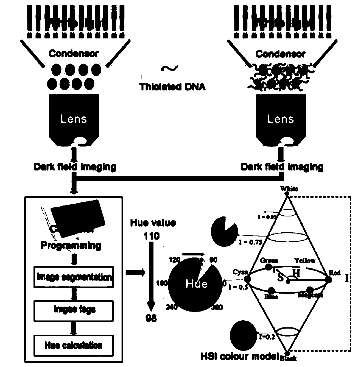
[0029] S1. Establish an HSI color model to form the basis for HSI analysis. The HSI color model requires that each color can be represented by three components: hue (H), saturation (S) and intensity (I);

[0030] The H component describes the color itself at an angle between [0, 360] degrees; 0 degrees represents red, 60 degrees represents yellow, 120 degrees represents green, 240 degrees represents blue, and 300 degrees represents magenta;

[0031] The S component represents the white pollution level, and its range is [0, 1];

[0032] The I component represents brightness information, and the range between [0,1] and 0 represents black, and 1 represents white;

[0033] The steps to build the model are as follows:

[0034] S1.1, use the DFM imaging system to measure the scattered light IDFMS of plasmonic n

Embodiment 2

[0041] Take the combination of sulfurized DNA and gold nanoparticles (AuNP) as an example to form the basis of HSI analysis:

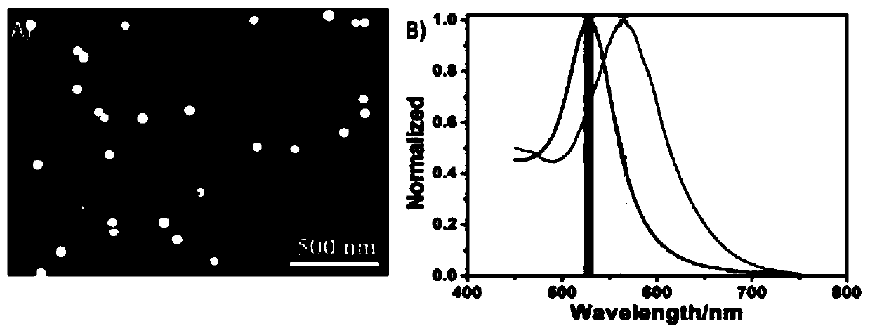
[0042] 1) The scattered light IDFMS of AuNP before and after sulfurized DNA attachment was measured by DFM imaging system;

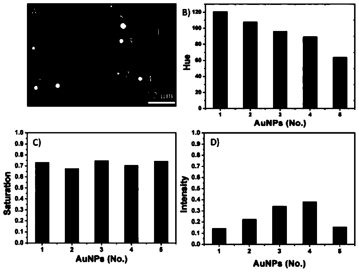
[0043]2) Through computer programming, the color spots in the obtained scattered light IDFMS are automatically converted into digital hue values ​​in the HSI color model; the algorithm of this program includes image segmentation, image labeling and hue calculation of each particle in the pixel; figure 1 It shows the change of the hue value of the scattered light color after the sulfurized DNA is attached to the surface of a single AuNP. After the sulfurized DNA is attached to the AuNP, the scattered light color of the single AuNP changes, and the corresponding hue value decreases;

[0044] 3) By encoding the scattered light color of the PNP with the HSI color system, the basis of the HSI analysis is formed;

[0045] Such as figur

Embodiment 3

[0051] To study the change of the color of light scattered by a single AuNP, to directly verify the possibility of using the HSI model to analyze the color of light scattered by a single AuNP, and to establish the relationship between the hue value of the standard spectral color and the shift of the scattering spectrum:

[0052] 1) Since the color change of the light scattered by a single AuNP is very sensitive to the surrounding medium, water, ethanol, 1-butanol, ethylene glycol (eg) and Soak AuNP in dimethyl sulfoxide (dmso);

[0053] 2) Through the above-mentioned computer programming, the hue value of the scattered light color of each AuNP in the DFM image can be automatically calculated.

[0054] The soaked AuNPs have different scattered light colors, gradually changing from green to yellow-green or yellow, where the red-shift of the scattered light occurs with the increase of Ri of the solvent, Figure 4 B is the obtained linear relationship between the hue value of indivi


PUM

PropertyMeasurementUnit
Average sizeaaaaaaaaaa

Abstract

The invention discloses a plasma nanoparticle dark field microimaging analysis method based on HSI color coding, and specifically relates to the field of quantitative chemistry and biochemical imaging. The method comprises the following specific analysis steps: S1, establishing an HSI color model, forming an HSI analysis basis, and the HSI color model requiring that each color can be represented by three components of hue H, saturation S and intensity I; S2, after obtaining an LSPR scattering signal of a single plasma nanoparticle, analyzing scattered light in the DFM image by using a hue value in the HSI system; and S3, perfecting the HSI color model. According to the method, the LSPR scattering signal of a single AuNP can be automatically obtained by encoding the pixels of the scatteredlight and adopting an HSI analysis method, HSI analysis is easier to implement, and the method is closer to a human visual system than RGB analysis. Besides, through computer programming, the hue value corresponding to each light spot in the DFM image can be automatically calculated and acquired, the precision is greatly improved, and personal errors are eliminated.

Description


Claims


Application Information

Patent Timeline
Owner SOUTHWEST UNIVERSITY
Why Eureka
Try Eureka