Method for culturing cell sphere by using self-assembled polypeptide derivative hydrogel, cell sphere and application of cell sphere

A technology for peptide derivatives and cell culture, applied in biochemical equipment and methods, epidermal cells/skin cells, tissue culture, etc., can solve the problems of low cell activity and complex preparation process of cell spheroids

Active Publication Date: 2022-03-22
NANKAI UNIV
View PDF3 Cites 0 Cited by
  • Summary
  • Abstract
  • Description
  • Claims
  • Application Information

AI Technical Summary

Benefits of technology

This technology allows researchers to efficiently create small spheres containing living tissue or cancerous areas that behave like real ones when placed into solution. These tiny particles will stay alive even after being treated with drugs because they maintain their original shape during storage without losing any properties such as chemicals or other substances from them. They may then help scientists study how different types of diseases develop more accurately than traditional methods.

Problems solved by technology

This patented technical problem addressed in this patents relates to producing stable and effective cell syroids without causing issues like poor viable culture conditions during manufacturing processes.

Method used


Image

  • Method for culturing cell sphere by using self-assembled polypeptide derivative hydrogel, cell sphere and application of cell sphere
  • Method for culturing cell sphere by using self-assembled polypeptide derivative hydrogel, cell sphere and application of cell sphere
  • Method for culturing cell sphere by using self-assembled polypeptide derivative hydrogel, cell sphere and application of cell sphere

Examples


Embodiment 1

[0051] Weigh Biotin- D 10 mg of F-YIGSR polypeptide derivative powder was placed in a glass vial, and 994.84 μL of 1×PBS (pH=7.4) and 5.16 μL of 1M Na were added 2 CO 3 solution, and mix well. Heat the vial with an alcohol lamp until the solution boils, then let it stand still, and after cooling to room temperature, the self-assembled polypeptide derivative hydrogel is obtained.

Embodiment 2

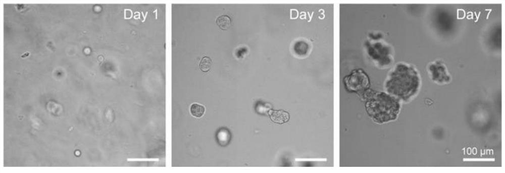
[0053] Take 37.5 μL of the self-assembled polypeptide derivative hydrogel obtained in Example 1, vortex and mix evenly with 12.5 μL of MCF-7 cell suspension containing 3000 cells, transfer to a 96-well plate, and put it into a cell culture incubator. After the cell gel mixture was placed in the incubator for 10 min, 100 μL of DDMEM (10% FBS) medium was added to its surface, and 50 μL of fresh complete medium was replaced every other day.

[0054] Record the growth of the cell spheroids, the results are as follows: figure 1 shown.

[0055] The results showed that the cell spheres could form normally and gradually increase in the 1-7 days; on the 7th day, the average cross-sectional area of ​​the cell spheres formed by MCF-7 was 4610 μm 2 .

Embodiment 3

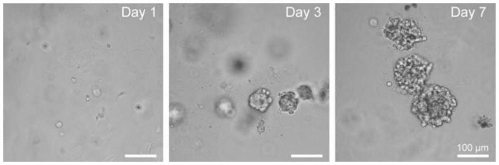
[0057] Take 37.5 μL of the self-assembled polypeptide derivative hydrogel obtained in Example 1, vortex and mix evenly with 12.5 μL of 4T1 cell suspension containing 3000 cells, transfer to a 96-well plate, and put it into a cell culture incubator. After the cell gel mixture was placed in the incubator for 12 minutes, 100 μL of RPMI1640 (10% FBS) medium was added to its surface, and 50 μL of fresh complete medium was replaced every other day.

[0058] Record the growth of the cell spheroids, the results are as follows: figure 2 shown.

[0059] The results showed that the cell spheres could form normally and gradually increase in the 1-7 days; on the 7th day, the average cross-sectional area of ​​the cell spheres formed by 4T1 was 11450 μm 2 .


PUM

PropertyMeasurementUnit
Average cross-sectional areaaaaaaaaaaa

Abstract

The invention provides a method for culturing a cell sphere by utilizing self-assembled polypeptide derivative hydrogel, the cell sphere and application of the cell sphere, and belongs to the technical field of biological materials. The culture process of the cell sphere comprises co-culture with the self-assembled polypeptide derivative hydrogel, and the structure of the self-assembled polypeptide derivative in the self-assembled polypeptide derivative hydrogel is Biotin-DPhe-X. The cell sphere provided by the invention overcomes the problems of complex cell sphere preparation process and low cell activity in the prior art, the cell sphere can be effectively obtained through co-culture of the self-assembled polypeptide derivative hydrogel and the cell suspension, the activity of the cell sphere is ensured, and when the used cells are stem cells, the cell dryness can be retained. The cell sphere can also be used for preparing organoids and tumors, and the tumors obtained through the cell sphere are similar to solid tumors in feature and can be used for drug screening.

Description


Claims


Application Information

Patent Timeline
Owner NANKAI UNIV
Why Eureka
Try Eureka