Medical image processing system, endoscope system, diagnosis support apparatus, and medical service support apparatus

a technology of medical image processing and endoscope, which is applied in the field of medical image processing system, endoscope system, diagnosis support apparatus, etc., can solve the problem of difficult to grasp the observation target on the white light image, and achieve the effect of reliably detecting a region and easy to grasp the observation targ

Active Publication Date: 2020-07-09
FUJIFILM CORP
View PDF0 Cites 0 Cited by
  • Summary
  • Abstract
  • Description
  • Claims
  • Application Information

AI Technical Summary

Benefits of technology

The technology described in this patent allows for reliable detection of areas that may be affected by changes in their appearance due to different operations being performed. This makes it easier for users to see what they are doing during these procedures.

Problems solved by technology

In this patent text describes how medical image processing systems use special lights for better visualization of small areas called lesions. These special lights have specific colors depending on their location within the body being examined. This allows users who don't know what they see when looking at them during surgery or other procedures to easily identify any abnormalities.

Method used


Image

  • Medical image processing system, endoscope system, diagnosis support apparatus, and medical service support apparatus
  • Medical image processing system, endoscope system, diagnosis support apparatus, and medical service support apparatus
  • Medical image processing system, endoscope system, diagnosis support apparatus, and medical service support apparatus

Examples


first embodiment

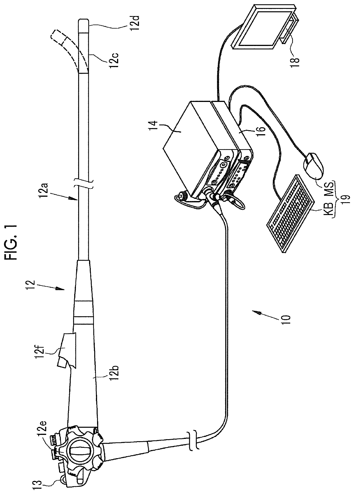
[0041]As shown in FIG. 1, an endoscope system 10 comprises an endoscope 12, a light source device 14, a processor device 16, a monitor 18, and a user interface 19. The endoscope 12 irradiates an observation target with illumination light, and images the observation target irradiated with the illumination light. The light source device 14 generates illumination light to be emitted to the observation target. The processor device 16 performs system control of the endoscope system 10, image processing, and the like. The monitor 18 is a display unit that displays an image output from the processor device 16. The user interface 19 is an input device for performing a setting input or the like with respect to the processor device 16 and the like, and is configured to include a keyboard KB, a mouse MS, and the like.

[0042]The endoscope 12 has an insertion part 12a that is to be inserted into a subject, an operation part 12b provided in a proximal end portion of the insertion part 12a, and a bend


PUM


Abstract

An imaging unit acquires a white light image by imaging an observation target being illuminated with white light, and acquires a specific light image by imaging the observation target being illuminated with specific light. A light source control unit controls the number of light emission frames of the white light and the number of light emission frames of the specific light in one light emission cycle according to at least one of a moving speed of the imaging unit, zoom information, or a light emission amount.

Description


Claims


Application Information

Patent Timeline
Owner FUJIFILM CORP
Why Eureka
Try Eureka