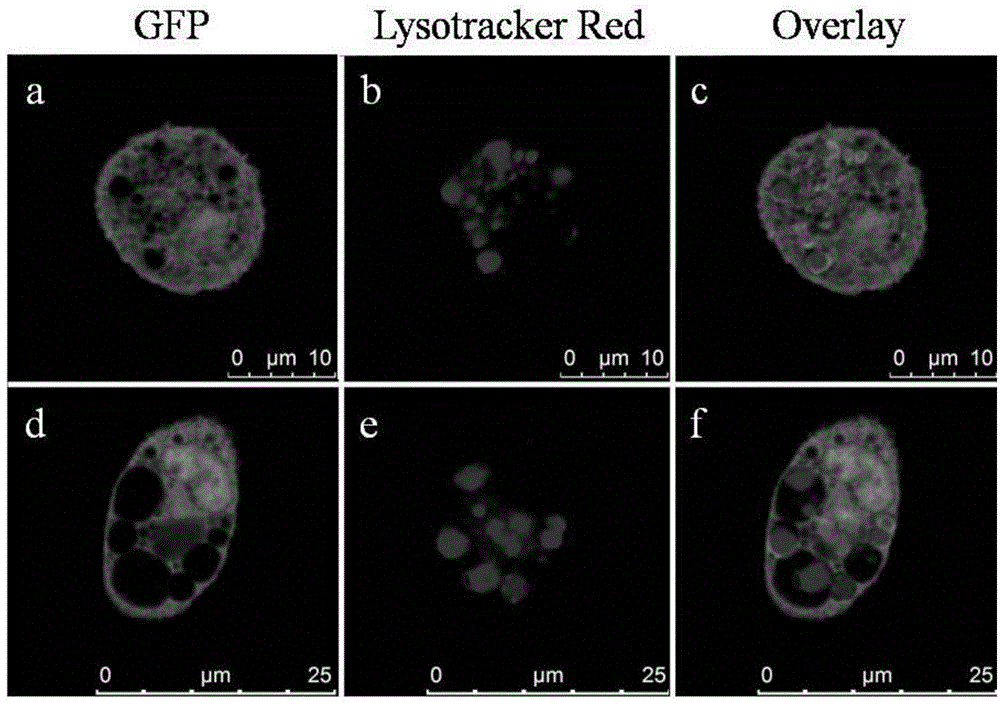
Method for observing morphology and functions of lysosomes by using transgenic macrophage expressing GFP or mutants thereof

A macrophage and transgenic technology, applied in the direction of biochemical equipment and methods, microbial measurement/testing, etc., can solve the problems of lysosomal marker omission, experimental error, unfavorable lysosomal function research, etc.

Inactive Publication Date: 2016-02-10
何向锋 +1
View PDF0 Cites 1 Cited by
  • Summary
  • Abstract
  • Description
  • Claims
  • Application Information

AI Technical Summary

Benefits of technology

This patented technology allows for better understanding of how cells break down organisms by observing their structure inside them. It involves introducing tiny particles called DNA into living tissues like bacteria which then use this small particle's properties to help study these processes more easily. By measuring specific characteristics such as size distribution, shape, color change, etc., we could get an idea about where certain parts were being broken up during cellular metabolism.

Problems solved by technology

This patents describes methods that use light waves to measure changes caused by certain substances inside live cells called chloroinospora lipids ("ChiL"). These techniques require strong basic conditions during analysis due to the presence of other compounds like lysolecan hydrochloride and necrosistinium sulfite within the cytoplasm of Chlamydia spp., causing interference when analyzed through transmitted electronic scanning optical instruments. Additionally, current procedures involve multiple steps involving chemical reactions between labeled molecules and target structures outside the body, leading to potential variations in pH levels throughout the sample being studied.

Method used


Image

  • Method for observing morphology and functions of lysosomes by using transgenic macrophage expressing GFP or mutants thereof
  • Method for observing morphology and functions of lysosomes by using transgenic macrophage expressing GFP or mutants thereof
  • Method for observing morphology and functions of lysosomes by using transgenic macrophage expressing GFP or mutants thereof

Examples


Embodiment Construction

[0037] The present invention can be better understood from the following examples. However, those skilled in the art can easily understand that the content described in the embodiments is only for illustrating the present invention, and should not and will not limit the present invention described in the claims.

[0038] 1. Materials

[0039] 1. Experimental Animals and Cells

[0040] Green fluorescent protein (GFP) transgenic mice were purchased from the Experimental Animal Center of Nantong University.

[0041] 2. Main reagents and instruments

[0042] Lysosome red fluorescent probe Lyso-TrackerRed, endoplasmic reticulum red fluorescent probe ER-TrackerRed and nucleus blue fluorescent probe DAPI were purchased from Beyontian Biotechnology Co., Ltd.; laser scanning confocal microscope (laserscanningconfocalmicroscope, LSCM) TCSSP5Ⅱ The type is the product of Leica Company of Germany; the special glass-bottom Petri dish for LSCM is the product of Wuxi Nice Biotechnology Co., L


PUM


Abstract

The invention discloses a method for observing morphology and functions of lysosomes by using transgenic macrophage expressing GFP or mutants thereof. The method includes steps of separating and cultivating transgenic macrophage suspension expressing GFP or mutants thereof; observing the separated and cultivated macrophage under laser scanning confocal microscopes. The invention further discloses application of the expression GFP or the transgenic macrophage suspension to observing the morphology and the functions of the lysosomes by the aid of the laser scanning confocal microscopes. The method and the application have the advantages that the lysosomes can be observed by using transgenic macrophage expressing GFP or mutants thereof, and accordingly the locations of the lysosomes can be clearly displayed without staining; the ultrastructure morphology, and the quantities and dynamic change of the lysosomes for expressing GFP or the transgenic macrophage of the mutants thereof can be clearly and visually observed by the aid of the laser scanning confocal microscopes; the quantities and the locations of primary lysosomes which are yet to start to realize digestion effects, corresponding substrates in the primary lysosomes and secondary lysosomes which realize digestion effects at the moment or completely realize the digestion effects further can be visually displayed if the substrates are labeled by red fluorescent probes.

Description


Claims


Application Information

Patent Timeline
Owner 何向锋
Why Eureka
Try Eureka