High-efficiency single-cell capture and rapid single-cell secretory protein detection platform and method
A secreted protein detection platform technology, applied in chemical instruments and methods, biochemical equipment and methods, tissue cell/virus culture devices, etc., can solve the problem of low sensitivity of single cell secreted protein detection and large volume of secreted protein detection cavity , low single cell capture efficiency and other problems, to achieve the effect of small size, high detection sensitivity and fast capture
- Summary
- Abstract
- Description
- Claims
- Application Information
AI Technical Summary
Problems solved by technology
Method used
Image


Examples
Embodiment 1
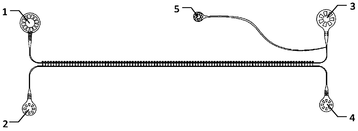
[0053] see Figure 1 to Figure 14 .High-efficiency single-cell capture and rapid single-cell secreted protein detection platform, including single-cell capture and droplet incubation microfluidic chip, secreted protein capture glass plate, chip fixture three parts; among them,
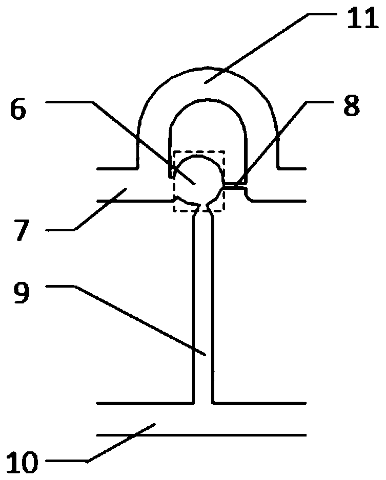
[0054] 1. See Figure 11 , the single cell capture and droplet incubation microfluidic chip includes: an isolation valve layer 23 on the upper layer and a single cell capture and droplet incubation layer 24 on the lower layer; the single cell capture and droplet incubation layer is connected end to end by multiple experimental units Each experimental unit includes a straight cell flow channel 7 located at the left and right ends of the cell flow channel. The two straight flow channels are connected by a U-shaped or arc-shaped cell flow channel 11. The left end of the cell capture chamber 6 is connected to the U Shaped or arc-shaped cell flow channel 11 is connected, the right end is connected with the ri
Embodiment 2
[0076] (1) Thermal bonding of single cell capture and droplet incubation layer PDMS chip, isolation valve layer PDMS chip
[0077]First use an upright microscope to align the single cell capture and droplet incubation layer with the isolation valve layer and stick them together, then place the chip in a 60-degree oven to heat for 20 minutes, then peel it off from the silicon wafer template, and Holes are punched at the entrance and exit of the chip channel, and the microfluidic chip for single cell capture and droplet incubation is completed.
[0078] (2) Modification of single cell capture and droplet incubation microfluidic chip
[0079] Use electronic fluoride solution 1720 to modify the channel of the chip. After modification, heat it in an oven for 1 hour to ensure that the solvent is completely evaporated, and then place the chip in PBS for boiling. After boiling, soak the chip in PBS and wait for the experiment. take out.
[0080] (3) Modification of single-cell secreted
Embodiment 3
[0084] Example 3 Modification of single-cell secreted protein capture glass plate
[0085] Fabrication of modified channel microfluidic PDMS chips, such as Figure 5 As shown, and punch holes at its entrance and exit; put the chip in absolute ethanol for 10 minutes, then put the chip in ultrapure water for 10 minutes, take it out and use nitrogen to dry the surface moisture, and put the chip in a clean place. Put it in a 80-degree oven and heat it for 30 minutes to dry; use adhesive tape to remove the residual impurities on the surface of the modified chip channel, attach the chip to the poly-lysine glass plate naturally, and place it in an 80-degree oven for heating Thermal bonding was performed for 2 hours; after it cooled down, BSA-FITC, IL-8 capture antibody, MCP-1 capture antibody, TNF-a capture antibody, and MIP-1b capture antibody were respectively passed through its 5 modification channels, 1.5 microliters each were used to modify the secreted protein capture antibody. T
PUM
| Property | Measurement | Unit |
|---|---|---|
| Length | aaaaa | aaaaa |
Abstract
Description
Claims
Application Information
 - R&D Engineer
- R&D Manager
- IP Professional
- Industry Leading Data Capabilities
- Powerful AI technology
- Patent DNA Extraction
Browse by: Latest US Patents, China's latest patents, Technical Efficacy Thesaurus, Application Domain, Technology Topic.
© 2024 PatSnap. All rights reserved.Legal|Privacy policy|Modern Slavery Act Transparency Statement|Sitemap