Lysosome fluorescent probe as well as preparation method and application thereof
A fluorescent probe and lysosome technology, applied in the field of fluorescent probes, can solve the problems of pH sensitivity and false positive signals, and achieve the effects of viscosity sensitivity, low toxicity and good photostability
- Summary
- Abstract
- Description
- Claims
- Application Information
AI Technical Summary
Problems solved by technology
Method used
Image

Examples
Embodiment 1
[0030] Synthesis of fluorescent probes
[0031] 1) Synthesis of benzindole iodoethanolate
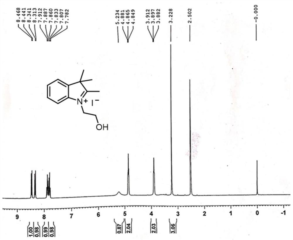
[0032] The compound 2,3,3-trimethyl-3H-indole (1.98 mL, 10 mmol) and 2-iodoethanol (1.72 mL, 10 mmol) were dissolved in 20 mL of pure ethanol solution and stirred in the flask for 1 hour. It was then refluxed for 8 h, cooled and filtered, and washed 3 times with anhydrous EtOH. After drying, 3.40 g of white solid was obtained, the yield: 92%, that is, compound II. And its hydrogen spectrum characterization, the results are as follows:
[0033] 1 H NMR (300 MHz, DMSO- d 6 ), δ (ppm): 8.45 (d, J = 8.1 Hz, 1H), 8.32 (d, J = 8.4 Hz, 1H), 7.89 (t, J = 7.8 Hz, 1H), 7.81 (t, J = 7.65 Hz, 1H), 5.23(s, 1H), 4.87 (t, J = 4.80 Hz, 2H), 3.90 (t, J = 4.50 Hz, 2H), 3.23 (s, 3H). Such as figure 1 shown.
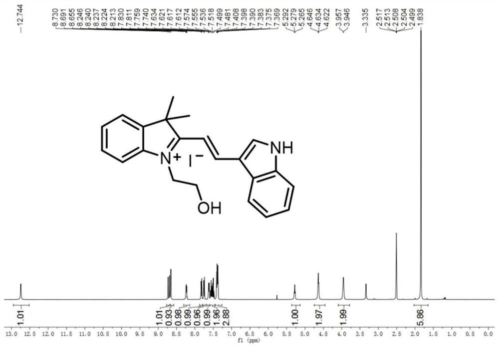
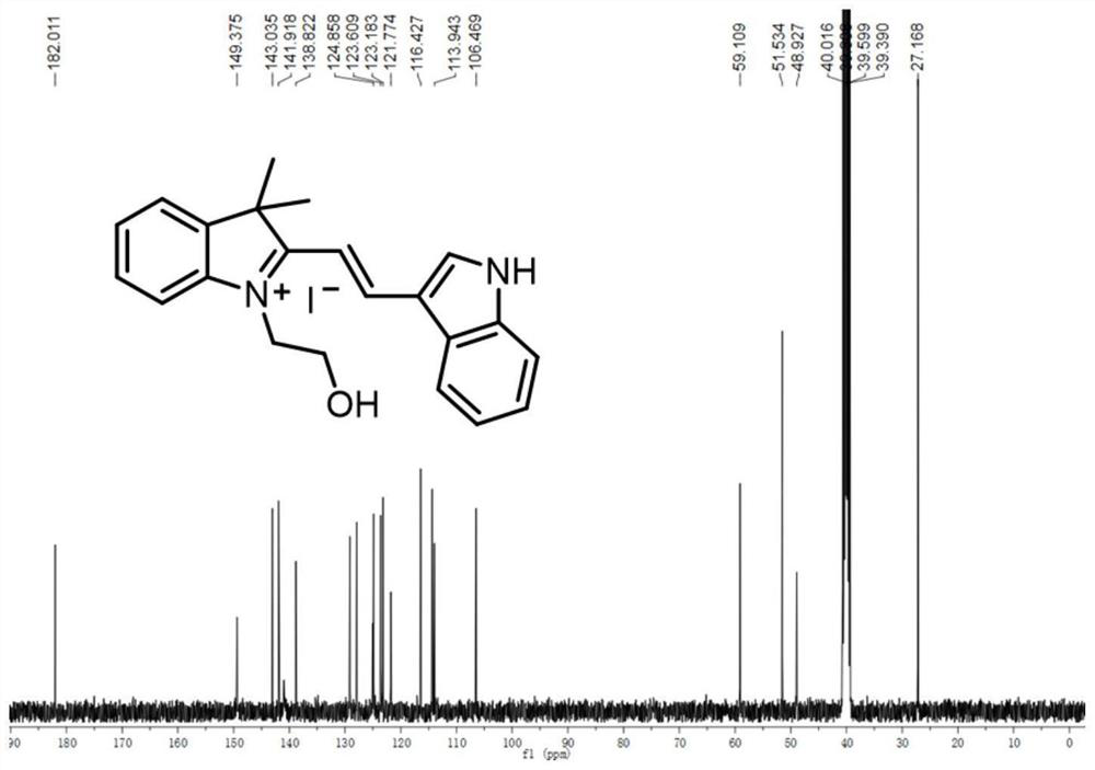
[0034] 2) Synthesis of fluorescent probe IVDI
[0035] Compound II (0.331 g, 1 mmol) and 1H-indole-2-carbaldehyde (0.145 g, 1 mmol) were dissolved in 20 mL of methanol, stirred in t
Embodiment 2
[0040] Toxicity Test of Probe IVDI - Standard MTT Method
[0041] HeLa cells growing in logarithmic phase were seeded in 96-well plates (about 1×10⁴ cells / well), and the wells were filled with cell-free medium as blank group. Place the seeded cells at 37°C, 5% CO 2 After incubation in the incubator for 24 h, 1, 2, 4, and 8 μM of IVDI were added to the wells as experimental groups. In addition, DMEM culture solution with a final concentration of 0.2% DMSO was added as a control group. Cells at 37°C, 5% CO 2 Incubate for 18 hours. MTT (5 mg / mL) was then added to each well. After incubation at 37°C for 4 h, 100 μL of DMSO was added. After incubation for another 20 minutes, the absorbance of each well at 490 nm was measured using a microplate reader, and the cytotoxicity experiment was repeated 4 times.
[0042] Cell viability can be calculated by the following formula:
[0043]
[0044] Among them, A sample is the absorbance of the experimental group, A c is the abso
Embodiment 3
[0046] IVDI's Photophysical Properties Test Experiment
[0047] Prepare test solutions containing 10 μM IVDI with solvents of different viscosities, and then prepare BR buffer solutions containing 2% DMSO at different pH values (pH= 4.0–9.0), and test their absorption and fluorescence emission spectra respectively. The results are shown in Figure 6 .
[0048] In panel (A), the probe IVDI has an absorption peak at 470 nm, and in panel (B), the fluorescence intensity of the probe is the strongest in glycerol and weaker in other low-viscosity solvents, indicating that The probe responds significantly to changes in viscosity. From panels (C) and (D), it can be seen that the probe IVDI barely responds to changes in pH. Experiments show that IVDI is a viscosity-sensitive probe that is not affected by pH changes. Probe IVDI can potentially image lysosomes with high fidelity and be used to observe changes in lysosome viscosity.
PUM
 Abstract
Description
Claims
Application Information
 - R&D Engineer
- R&D Manager
- IP Professional
- Industry Leading Data Capabilities
- Powerful AI technology
- Patent DNA Extraction
Browse by: Latest US Patents, China's latest patents, Technical Efficacy Thesaurus, Application Domain, Technology Topic.
© 2024 PatSnap. All rights reserved.Legal|Privacy policy|Modern Slavery Act Transparency Statement|Sitemap