Methods for diagnosing endometriosis
- Summary
- Abstract
- Description
- Claims
- Application Information
AI Technical Summary
Benefits of technology
Problems solved by technology
Method used
Image

Examples
example 1
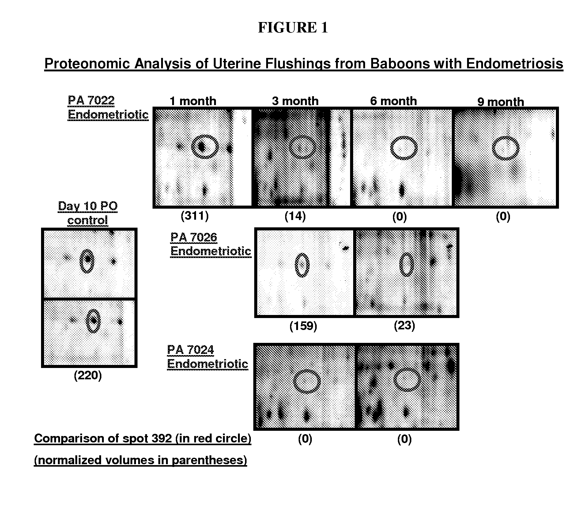
Proteonomic Analysis of Uterine Flushings
[0101]Uterine flushings were evaluated from control baboons and those with endometriosis to determine if changes would be evident that would be reflective of the disease condition. Uterine flushings were obtained at day 10 post ovulation (PO) from baboons with endometriosis (n=3) 1, 3, 6, 9, 12, and 15 months during disease progression. Control flushings were obtained from control animals (n=4) at day 10 PO. Seventy micrograms of protein from each control baboon flush (n=4) was labeled with Cy3; 70 μg of flush protein from baboons with endometriosis (n=3) was labeled with Cy5; the 70 μg of the control flushing that were pooled to serve as a reference standard was labeled with Cy2. The samples were subjected to 2-dimensional (2-D) gel electrophoresis. The first dimension was a pH 3-10 non-linear IPG strip. The second dimension was a 12.5% tris-glycine polyacrylamide gel. Following the 2-D separation, con-imaging of Cy2, Cy3, and Cy5 labeled prote
example 2
Human Protocol
[0103]The study will target women undergoing diagnostic laparoscopy for the investigation of pelvic pain or multiparity. On the day of her surgery, the consenting subject will undergo blood sampling (5 ml), cervical secretion collection (with a Dacron swab), a uterine flushing with 15 ml of sterile lactate ringers and an endometrial biopsy. The surgery will be performed while the subject is under anesthesia. During the laparoscopy, confirmation and staging of endometriosis will be performed using the revised ASRM classification scale, and the amount of endometriosis removed / percent of remaining disease will be estimated by the surgeon. Whenever possible, laparoscopic photographs will be taken to confirm the scoring process.
[0104]A second group of subjects with the diagnosis of multiparity, who are undergoing laparoscopic tubal ligation, will be used as controls. These subjects will undergo the same type of sample collections and will also be examined to rule out the pre
example 3
[0110]ELISA Analysis
[0111]Enzyme-linked immunosorbent assay analysis will be performed to detect and quantify the concentration of the β-subunit of fibrinogen in samples. A sandwich ELISA method will make use of anti-β subunit antibodies (capture antibodies), which may be noncovalently adsorbed (“coated”—primarily as a result of hydrophobic interactions) onto plastic microwell plates. After plate washings, the immobilized antibodies may specifically capture soluble β-subunit proteins present in samples that were applied to the plate. After washing away unbound material, the captured proteins are detected by a label-conjugated anti-β subunit antibodies (for example, biotin-labeled detection antibodies) followed by an enzyme-labeled avidin or streptavidin stage. Following the addition of a chromogenic substrate, the level of colored product generated by the bound, enzyme-linked detection reagents may be measured spectrophotometrically using an ELISA-plate reader at an appropriate optical
PUM
| Property | Measurement | Unit |
|---|---|---|
| Time | aaaaa | aaaaa |
| Time | aaaaa | aaaaa |
| Time | aaaaa | aaaaa |
Abstract
Description
Claims
Application Information
 - R&D Engineer
- R&D Manager
- IP Professional
- Industry Leading Data Capabilities
- Powerful AI technology
- Patent DNA Extraction
Browse by: Latest US Patents, China's latest patents, Technical Efficacy Thesaurus, Application Domain, Technology Topic.
© 2024 PatSnap. All rights reserved.Legal|Privacy policy|Modern Slavery Act Transparency Statement|Sitemap