Application of micelle as photosensitizer formed by amphiphilic molecules and Ag2S quantum dots
A technology of amphiphilic molecules and quantum dots, applied in the field of nanomaterials, can solve problems such as damage, necrosis, and phototoxicity
- Summary
- Abstract
- Description
- Claims
- Application Information
AI Technical Summary
Problems solved by technology
Method used
Image

Examples
Embodiment 1
[0036] Example 1: Test at the probe level to generate singlet oxygen after light excitation of silver sulfide after water transfer
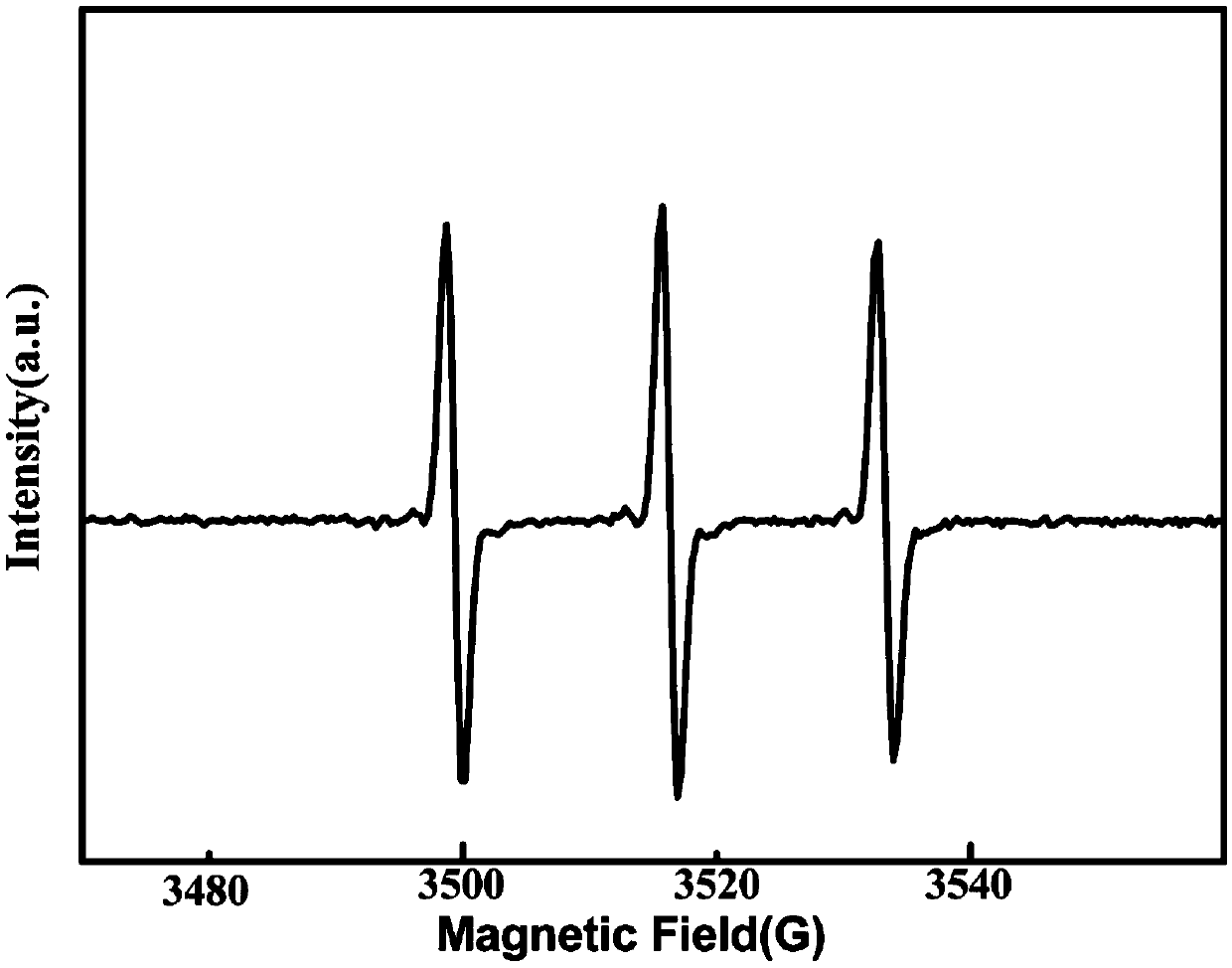
[0037] The use of singlet oxygen will combine with the capture agent TEMP to generate TEMPO, and TEMPO has a unique triple resonance signal peak. The Bruker electron spin resonance spectrometer was used as the detection system, the MDL-III-808-2.5W laser was used as the excitation light source, and the spectral data were collected by the control program.
[0038] Test method for singlet oxygen produced by near-infrared silver sulfide quantum dots after water transfer as a new photosensitizer probe:
[0039] Mix the silver sulfide (15μg / mL) after water transfer with the singlet oxygen scavenger TEMP (2,2,6,6-tetramethylpiperidine), and use the MDL-III-808-2.5W laser at 1.0w / cm 2 Under light-induced excitation for 10 min, the signal was detected on a Bruker electron spin resonance spectrometer.
[0040] The result is as figure 1 shown, from fi
Embodiment 2
[0041] Example 2: Detecting the change of singlet oxygen generation over time at the probe level
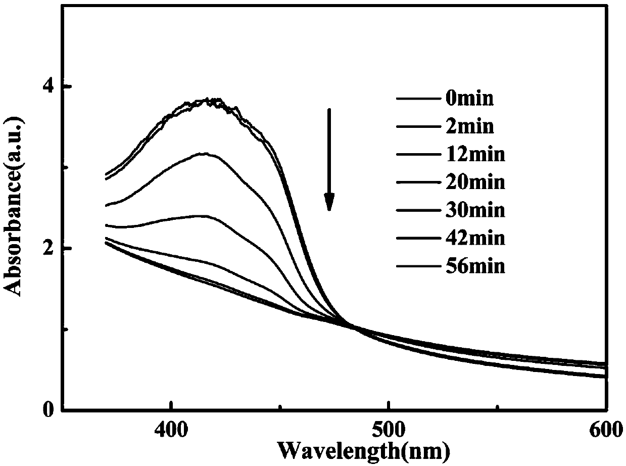
[0042] DPBF is one of the most active singlet oxygen scavenger known, and singlet oxygen will attack the furan ring in the DPBF structure to open it, resulting in a change in the absorption of DPBF at 410nm. Therefore, by co-incubating DPBF with silver sulfide after water transfer, after laser irradiation for different times, the absorption spectrum was collected on a UV-2550 ultraviolet-visible spectrophotometer.
[0043] The test method for the change of singlet oxygen produced by the near-infrared silver sulfide quantum dots as a new photosensitizer probe over time after water transfer is as follows:
[0044] Take 2.5mL of silver sulfide (15μg / mL) after water transfer into a quartz cuvette, add 100μL of DPBF (1,3-diphenylisobenzofuran), and pass through MDL-III-808-2.5W laser 1.0w / cm 2 After laser induction for different times (0, 2, 12, 20, 30, 42 and 56 min), the spectra
Embodiment 3
[0046] Example 3: Detection of singlet oxygen production at the cellular level
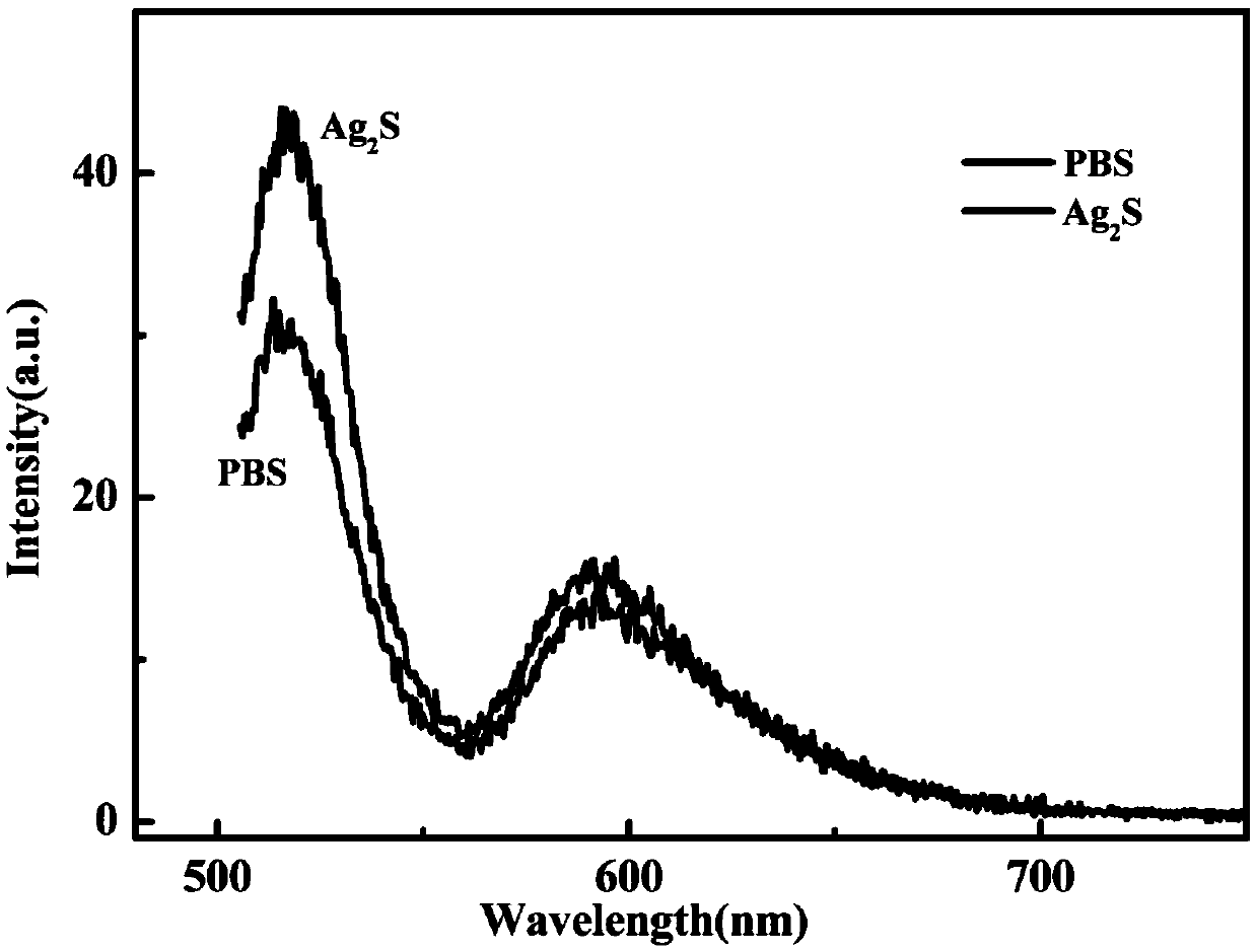
[0047] DCFH-DA is known as an active oxygen fluorescence detection kit. When DCFH-DA diffuses into the cell, it will deacetylate and become non-fluorescent DCFH, and DCFH can be rapidly oxidized by reactive oxygen species to DCF with high-intensity fluorescence, and the fluorescence intensity is related to the ROS content in the cell fluid. proportional. Therefore, after HeLa cells were co-incubated with the probe after water transfer, and after the MDL-III-808-2.5W laser was excited by the light induction, the emission of different fluorescence spectra was recorded by the QE6500 fluorescence spectrometer, and different methods were observed by the fluorescence microscope. Fluorescence imaging of processed cells was performed to detect the ability of the probe to induce singlet oxygen production.
[0048] The near-infrared silver sulfide quantum dot after water transfer is used as a new photosensit
PUM
| Property | Measurement | Unit |
|---|---|---|
| Wavelength | aaaaa | aaaaa |
| Power density | aaaaa | aaaaa |
| Diameter | aaaaa | aaaaa |
Abstract
Description
Claims
Application Information
 - R&D Engineer
- R&D Manager
- IP Professional
- Industry Leading Data Capabilities
- Powerful AI technology
- Patent DNA Extraction
Browse by: Latest US Patents, China's latest patents, Technical Efficacy Thesaurus, Application Domain, Technology Topic.
© 2024 PatSnap. All rights reserved.Legal|Privacy policy|Modern Slavery Act Transparency Statement|Sitemap